Pelvic Bone
Table of Contents
Introduction
The pelvic bone, also known as the hip bone or innominate bone, is a large, sturdy bone located in the lower part of the trunk. It is a complex structure that consists of three fused bones: the ilium, ischium, and pubis.
The pelvic bone plays a crucial role in supporting the weight of the upper body and connecting the spine to the lower limbs. It forms the pelvis, which protects and houses various organs including the reproductive organs, bladder, and rectum. Additionally, it serves as an attachment point for several muscles involved in the movement and stability of the hip joint.
The pelvic bone has distinct features that differ between males and females. In females, the pelvis is generally wider and shallower to accommodate childbirth, while in males, it is narrower and more robust to support greater muscle mass and stability.
Injuries to the pelvic bone can occur due to trauma, such as falls or car accidents, resulting in fractures or dislocations. These injuries can be serious and may require medical intervention.
Overall, the pelvic bone is a vital structure in the human body, providing support, and protection, and facilitating movement and stability.
Parts of pelvic
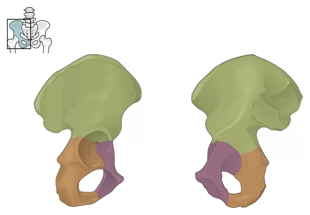
The pelvis bone is composed of three main parts: the ilium, ischium, and pubis.

- Ilium: The ilium is the largest and uppermost part of the pelvis bone. It is located on each side of the body and forms the broad, curved upper section of the pelvis. The ilium consists of a body and two wings called alae. The body of the ilium connects to the sacrum, a triangular bone at the base of the spine, forming the sacroiliac joint. The alae of the ilium project outward and provide attachment points for muscles and ligaments.
- Ischium: The ischium is located at the lower and posterior part of the pelvis bone. It is commonly referred to as the “sit bone” as it bears weight when sitting. The ischium consists of a body and a ramus. The body of the ischium forms the lower part of the acetabulum, which is a socket that connects with the femur (thigh bone) to form the hip joint. The ramus extends from the body of the ischium towards the pubis bone, where it fuses with it.
- Pubis: The pubis is located at the front part of the pelvis bone. It consists of a body and two rami (plural of ramus). The body of the pubis joins with the body of the opposite pubis bone at the midline, forming a joint called the pubic symphysis. This joint allows for slight movement and flexibility during activities such as walking or childbirth. The rami of the pubis extend from the body towards the ischium and fuse with it, completing the pelvic ring.
These three parts of the pelvis bone are connected by fibrous joints known as sutures. During development, these bones are separate, but they gradually fuse together as a person grows. The pelvis bone, along with the sacrum and coccyx (tailbone), forms the bony pelvis, which provides support for the abdominal organs and helps transmit weight from the upper body to the lower limbs.
Joint articulation
The pelvic joint articulation refers to the connections between the three main parts of the pelvis bone: the ilium, ischium, and pubis. These joints allow for movement and flexibility in the pelvis, while also providing stability and support.
- Sacroiliac Joint: The sacroiliac joint is formed by the connection between the body of the ilium and the sacrum. The sacrum is a triangular bone at the base of the spine that fits between the two iliac bones. The joint is supported by strong ligaments that help stabilize the pelvis. The sacroiliac joint has limited mobility, as its primary function is to transmit weight from the upper body to the lower limbs.
- Pubic Symphysis: The pubic symphysis is a joint located at the midline of the pelvis, where the bodies of the two pubic bones meet. It is a cartilaginous joint, meaning it is connected by fibrocartilage rather than a true joint cavity. The fibrocartilage allows for slight movement and flexibility during activities such as walking or childbirth. The pubic symphysis is reinforced by ligaments that help maintain stability.
In addition to these joints, there are other articulations within the pelvis that contribute to its overall function:
- Acetabulofemoral Joint: This is the hip joint, where the acetabulum of the pelvis articulates with the head of the femur (thigh bone). It is a ball-and-socket joint that allows for a wide range of movement, including flexion, extension, abduction, adduction, rotation, and circumduction.
- Sacrococcygeal Joint: This joint connects the sacrum to the coccyx (tailbone) at the base of the spine. It is a small joint with limited mobility.
- Sacrotuberous and Sacrospinous Ligaments: These ligaments connect the sacrum to the ischial tuberosity (part of the ischium) and the spine of the ischium, respectively. They help provide stability to the pelvis and support the weight-bearing function.
Overall, the joint articulations of the pelvis allow for a combination of stability and mobility, enabling various movements such as walking, running, sitting, and childbirth. These joints work together to support the body’s weight, transmit forces, and accommodate the changing needs of different activities.
Functions of the pelvic bone
The pelvic bone, also known as the hip bone or innominate bone, is a large, irregularly shaped bone that forms part of the pelvis. It is created of three fused bones which are the ilium, ischium, and pubis. The functions of the pelvic bone are numerous and vital for various aspects of human anatomy and physiology. these are some of the pelvic bone’s important functions:
- Support and Stability: The pelvic bone provides support and stability to the spine, connecting the vertebral column to the lower limbs. It acts as a strong anchor for the weight-bearing structures of the body, such as the spine, pelvis, and lower extremities.
- Protection: The pelvic bone forms a protective enclosure for several vital organs located within the pelvic cavity. These include the reproductive organs (uterus, ovaries, and testes), the urinary bladder, and parts of the digestive system (rectum).
- Attachment Site for Muscles: Numerous muscles attach to the pelvic bone, playing essential roles in movement, posture, and maintaining balance. These muscles include the gluteal muscles (buttocks), hip flexors and extensors, adductors (inner thigh muscles), and abdominal muscles.
- Weight Distribution: The pelvic bone helps distribute the weight of the upper body to the lower limbs during standing, walking, running, and other weight-bearing activities. It transfers forces generated by the legs to the vertebral column, reducing stress on the lower back.
- Childbirth: In females, the pelvic bone plays a crucial role in childbirth. Its shape and structure allow for the passage of the baby through the birth canal during delivery. The pelvic cavity widens during pregnancy to accommodate the growing fetus.
- Blood Cell Production: The inner part of the pelvic bone contains red bone marrow, which is responsible for producing red blood cells, white blood cells, and platelets. This process is known as hematopoiesis and is vital for maintaining a healthy blood supply.
- Joint Function: The pelvic bone forms several joints, including the sacroiliac joints with the sacrum and the pubic symphysis joint between the two pubic bones. These joints allow for limited movement, primarily for shock absorption during activities such as walking or running.
- Balance and Posture: The pelvic bone, along with its associated muscles, ligaments, and joints, contributes to maintaining balance and proper posture. It provides a stable base for the spine, allowing for efficient movement and preventing excessive strain on the back.
In summary, the pelvic bone serves multiple functions, including support, protection, muscle attachment, weight distribution, childbirth facilitation, blood cell production, joint function, and balance/posture maintenance. Its complex structure and role make it an essential component of the human skeletal system.
Pelvic floor muscles

The pelvic floor muscles are a group of muscles that create a supportive hammock-like structure at the bottom of the pelvis. They span from the pubic bone in the front to the coccyx (tailbone) in the back and from one sitting bone to the other. These muscles play a crucial role in supporting the pelvic organs, including the bladder, uterus (in females), and rectum, as well as maintaining continence (control over urination and bowel movements).
The main muscles of the pelvic floor involve the levator ani and coccygeus muscles.
Levator Ani: This is a group of muscles that form the pelvic floor and attach to various points on the pelvis, including the pubic bone, ischial spine, and coccyx. They support the pelvic organs, assist in maintaining continence, and play a role in sexual function.
Coccygeus: This muscle is located at the back of the pelvis and attaches to the ischial spine and coccyx. It also contributes to pelvic floor support.
These muscles have various attachments to different points on the pelvis, including the pubic bone, ischial spine, and coccyx.
The pelvic floor muscles have some important functions:
- Support: They provide support to the pelvic organs, helping to keep them in their proper position. This is particularly important during activities such as coughing, sneezing, laughing, or lifting heavy objects, which can put pressure on the pelvic organs.
- Continence: The pelvic floor muscles help maintain control over urination and bowel movements. When these muscles are strong and coordinated, they can contract and relax at the appropriate times to allow for voluntary control over the release of urine and feces.
- Sexual function: The pelvic floor muscles play a role in sexual function by contributing to arousal and orgasm. They also help with vaginal tone and support during intercourse.
- Stability: The pelvic floor muscles work in conjunction with other core muscles to provide stability to the pelvis and spine. They help maintain proper alignment and posture, which can prevent issues such as lower back pain.
Weakness or dysfunction in the pelvic floor muscles can lead to various problems, including urinary or fecal incontinence, pelvic organ prolapse (when one or more of the pelvic organs descend into the vagina), and sexual dysfunction. Factors such as pregnancy, childbirth, aging, obesity, chronic coughing, and certain medical conditions can contribute to pelvic floor muscle weakness.
Exercises known as Kegel exercises are commonly recommended to strengthen the pelvic floor muscles. These exercises involve contracting and relaxing the muscles in a specific way to improve their strength and coordination. Pelvic floor physical therapy may also be recommended for individuals with more severe pelvic floor dysfunction.
It’s important to note that everyone’s pelvic floor muscles are unique, and it may be beneficial to consult with a healthcare professional, such as a physical therapist or gynecologist, for personalized advice and guidance on maintaining pelvic floor health.
Ligaments
The pelvic bones are attached to various ligaments that help provide stability and support to the pelvic region. These ligaments play a crucial role in maintaining the integrity of the pelvic floor and its surrounding structures. Here are some of the main ligaments that attach to the pelvic bones:
- Sacroiliac Ligaments: The sacroiliac (SI) ligaments connect the sacrum (the triangular bone at the base of the spine) to the ilium (the largest bone of the pelvis). There are three main SI ligaments: the anterior sacroiliac ligament, the interosseous sacroiliac ligament, and the posterior sacroiliac ligament. These ligaments provide stability to the sacroiliac joint and help transmit forces between the spine and lower extremities.
- Sacrotuberous Ligament: The sacrotuberous ligament extends from the sacrum to the ischial tuberosity, which is a bony prominence on the bottom part of the pelvis. This ligament helps support the weight of the body when sitting and provides stability to the sacrum.
- Sacrospinous Ligament: The sacrospinous ligament is located deep within the pelvis and connects the sacrum to the ischial spine, which is another bony prominence on the pelvis. This ligament helps stabilize the sacrum and supports the pelvic organs.
- Pubic Symphysis: The pubic symphysis is a joint located at the front of the pelvis, where the two pubic bones meet. It is stabilized by a fibrocartilaginous disc and several ligaments, including the superior pubic ligament, inferior pubic ligament, and arcuate pubic ligament. These ligaments help maintain the stability and alignment of the pubic symphysis.
- Iliofemoral Ligament: The iliofemoral ligament is located in the front of the hip joint and connects the ilium to the femur (thigh bone). It is one of the strongest ligaments in the body and helps prevent excessive extension of the hip joint.
- Sacroiliolumbar Ligament: The sacroiliolumbar ligament connects the transverse process of the fifth lumbar vertebra (lower back) to the ileum. It provides stability to the sacroiliac joint and helps transmit forces between the spine and pelvis.
These ligaments work together to provide stability, support, and proper alignment of the pelvic bones. They play a crucial role in maintaining pelvic floor health and preventing issues such as pelvic organ prolapse or instability.
Blood supply
blood supply The blood supply to the pelvic region is crucial for providing oxygen and nutrients to the pelvic organs, muscles, and other structures. The blood vessels that supply the pelvis include arteries and veins.
Arteries:
- Internal Iliac Arteries: The internal iliac arteries are the main arteries that supply blood to the pelvis. They arise from the common iliac arteries, which are the large arteries that branch off from the abdominal aorta. The internal iliac arteries divide into several branches that supply different regions of the pelvis.
- Anterior Division Branches: These branches include the umbilical artery (which becomes the medial umbilical ligament in adults), superior vesical arteries (supplying the urinary bladder), inferior vesical artery (supplying the prostate in males and the uterus in females), middle rectal artery (supplying the rectum), and obturator artery (supplying the muscles of the inner thigh).
- Posterior Division Branches: These branches include the iliolumbar artery (supplying the muscles and bones of the lower back), lateral sacral arteries (supplying the sacrum and coccyx), and superior gluteal artery (supplying the gluteal muscles).
- Median Sacral Artery: The median sacral artery arises from the abdominal aorta just above its bifurcation into the common iliac arteries. It descends along the midline of the sacrum, supplying blood to the sacrum and coccyx.
Veins:
The veins that drain blood from the pelvic region include:
- Internal Iliac Veins: The internal iliac veins collect blood from the pelvic organs, muscles, and other structures. They eventually connect with the external iliac veins to create the common iliac veins.
- Median Sacral Vein: The median sacral vein accompanies the median sacral artery and drains blood from the sacrum and coccyx. It usually drains into the internal iliac veins.
- Other Veins: The pelvic region also contains various smaller veins that drain blood from specific structures. For example, the vesical venous plexus drains blood from the urinary bladder, and the rectal venous plexus drains blood from the rectum.
The blood supply to the pelvic region is important for maintaining the health and function of the pelvic organs and structures. It ensures proper oxygenation and nutrient delivery, as well as the removal of waste products. Disruption of the blood supply can lead to various issues, such as ischemia (lack of blood flow), thrombosis (blood clot formation), or hemorrhage (excessive bleeding).
Conditions that affect pelvic
There are several conditions that can affect the blood supply to the pelvic region, leading to various health issues. Some of these conditions include:

- Pelvic Congestion Syndrome: This condition occurs when there is increased pressure in the veins of the pelvis, causing them to become enlarged and dilated. It can lead to chronic pelvic pain, discomfort, and a feeling of fullness in the pelvis. Pelvic congestion syndrome is more common in women and can be caused by factors such as pregnancy, hormonal imbalances, or anatomical abnormalities.
- Pelvic Inflammatory Disease (PID): The uterus, fallopian tubes, and ovaries are all affected by the infectious condition known as pelvic inflammatory disease (PID). Chlamydia and gonorrhoea are two sexually transmitted illnesses that frequently cause it. PID can result in inflammation and scarring of the pelvic organs, leading to chronic pelvic pain, infertility, and an increased risk of ectopic pregnancy.
- Endometriosis: Endometriosis is a condition where the tissue that normally lines the uterus (endometrium) grows outside of the uterus, such as on the ovaries, fallopian tubes, or pelvic lining. This abnormal tissue growth can cause inflammation, scarring, and adhesions in the pelvis. Endometriosis often leads to severe pelvic pain, painful periods, and fertility problems.
- Pelvic Arterial Occlusive Disease: This condition occurs when there is a blockage or narrowing of the arteries that supply blood to the pelvis. It can be caused by atherosclerosis (build-up of plaque in the arteries), arterial thrombosis (blood clot formation), or arterial dissection (tearing of the artery wall). Pelvic arterial occlusive disease can result in reduced blood flow to the pelvic organs, leading to symptoms such as pain, numbness, or erectile dysfunction in men.
- Pelvic Venous Thrombosis: This condition involves the formation of blood clots in the veins of the pelvis. It can be caused by factors such as pregnancy, hormonal changes, or prolonged immobilization. Pelvic venous thrombosis can lead to pain, swelling, and discomfort in the pelvis, as well as potential complications such as pulmonary embolism (when a blood clot travels to the lungs).
- Pelvic Trauma: Trauma to the pelvis, such as from a car accident or a fall, can cause damage to the blood vessels in the pelvic region. This can result in bleeding, hematoma formation (collection of blood), or disruption of blood flow to the pelvic organs. Pelvic trauma can lead to severe pain, internal bleeding, and potentially life-threatening complications.
It is important to note that these conditions can have overlapping symptoms and may require medical evaluation and diagnosis by a healthcare professional. Treatment options for these conditions vary depending on the underlying cause and severity and may include medications, surgical interventions, or lifestyle modifications.
Exercise for pelvic
Exercise for the pelvic bone focuses on strengthening the muscles and improving the stability and mobility of the pelvic region. Here are some activities that can be beneficial for the pelvic bone:

Pelvic Tilts: Lie on your back with your knees bent and feet level on the ground. Slowly tilt your pelvis forward, pressing your lower back into the floor. maintain for some seconds, after that tilt your pelvis backward side, arching your lower back slightly. do it again this movement 10-15 times, focusing on engaging the muscles around the pelvic bone.
Bridge Pose: Lie on your back with your knees must be bent and feet should be level on the ground. raise your hips off the ground, and form a straight line from your knees to your shoulders. Squeeze your glutes and engage your core as you maintain this posture for some seconds. Lower your hips back down and do it again for 10-15 repetitions.
Clamshells: Lie on your side with knees bent and legs placed on each other. Keeping your feet together, lift your top knee while keeping your feet touching. Lower your knee back down and repeat for 10-15 repetitions on each side. This exercise targets the muscles around the hips and pelvis.
Squats: Stand with feet hip-width apart and toes pointing slightly outward. Bend your knees and reduce your hips as if sitting back in a chair. Keep your chest raised and your weight in your heels. get Pushed with your heels to get back to the initiating position. Repeat for 10-15 repetitions. Squats help strengthen the muscles in the thighs, hips, and pelvis.

Lunges: Stand with feet hip-width apart. Take a step forward with one foot and lower your body down until both knees are bent at a 90-degree angle. get pushed with the front heel to get back to the initial position. Do it again on the other side and continue the other side for 10-15 repetitions. Lunges target the muscles in the hips and pelvis, as well as the thighs.
Kegel Exercises: Kegel exercises include contracting and releasing the pelvic floor muscles. To perform a Kegel exercise, imagine stopping the flow of urine midstream. Squeeze and hold those muscles for a few seconds, then release. Repeat this contraction and relaxation pattern for 10-15 repetitions. Kegel exercises help strengthen the muscles that support the pelvic bone and can improve pelvic floor function.
It is important to start with gentle exercises and gradually increase intensity as tolerated. If you have any specific pelvic conditions or concerns, it is recommended to consult with a physiotherapist who specializes in pelvic health to ensure you are performing the exercises correctly and safely.
Summary
The pelvis bone, also known as the pelvic girdle or hip bone, is a large, sturdy bone located in the lower part of the body. It consists of several fused bones, including the ilium, ischium, and pubis. The pelvis bone forms a ring-like structure that connects the spine to the lower limbs and provides support for the body’s weight. It also protects and houses various organs, such as the reproductive organs, bladder, and rectum. The pelvis bone plays a crucial role in supporting the body during movement and providing stability for the lower limbs.
FAQ
The pelvic bone plays a crucial role in supporting the weight of the upper body and connecting the spine to the lower limbs. It forms the pelvis, which protects and houses various organs including the reproductive organs, bladder, and rectum. Additionally, it serves as an attachment point for several muscles involved in the movement and stability of the hip joint.
The pelvic bone has distinct features that differ between males and females. In females, the pelvis is generally wider and shallower to accommodate childbirth, while in males, it is narrower and more robust to support greater muscle mass and stability.
Injuries to the pelvic bone can occur due to trauma, such as falls or car accidents, resulting in fractures or dislocations.
Injuries to the pelvic bone can be serious because they can cause significant pain, difficulty walking or moving, and may even damage nearby organs. They often require medical intervention for proper treatment and healing.
The pelvic bone is a vital structure in the human body as it provides support, and protection, and facilitates movement and stability. It supports the weight of the upper body, connects the spine to the lower limbs, protects organs, and serves as an attachment point for muscles involved in hip joint movement and stability.