Lumbrical Muscle of the Hand
Table of Contents
Description
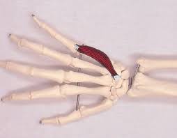
The lumbrical muscles are a group of four small intrinsic muscles located in the hand. They are named for their similarity to small earthworms or “lumbrical.” These muscles are responsible for various movements and functions within the hand.
Because of their worm-like impression (lumbrical means earthworm in Latin), the 4 short intrinsic muscles of the hand that are located in the middle of the metacarpal bones and deep into the palmar fascia are directed as the lumbrical muscles. The lumbrical muscles, along with the dorsal and palmar interossei, are among the hand’s short muscles.
At the MCP and IP joints, the lumbrical muscles of the hand flex and stretch the fingers and thumbs. Many hand functions, namely gripping motions, depend on them.
Structure of the Lumbrical muscles
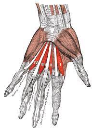
The lubricants are four tiny, worm-like muscles in each hand. The fact that these muscles aren’t attached to bone separates them. Rather, lumbricals attach distally to the extensor expansions and proximally to the flexor digitorum profundus tendons.
Origin of the first number of head
It originates on the radial side of the flexor digitorum profundus’s most outspread ligament (in comparison to the pointer finger).
Insertion
After traveling posteriorly along the radial side of the index finger, it inserts on the extensor expansion at the metacarpophalangeal joint.
Origin of the second number of head
It is originate from the 2nd-most radial tendon of the flexor digitorum profundus, which is situated on the radial side of the middle finger.
Insertion
It attaches to the extensor contraction around the metacarpophalangeal joint and goes on posteriorly along the radial side of the middle finger.
Origin of the third number of head
When compared to the ring finger, one head originates on the radial side of the flexor digitorum profundus ligament, while the other originates on the ulnar side of the ligament.
Insertion
The muscle travels posteriorly down to the radial side of the ring finger to insert its extensor expansion.
Origin of the fourth number of head
One head forms on the radial side of the flexor digitorum profundus tendon in the little finger and the other on the ulnar side of the tendon in the ring finger.
Insertion
The muscle goes posteriorly down the radial side of the little finger to insert its extensor expansion.
Innervation
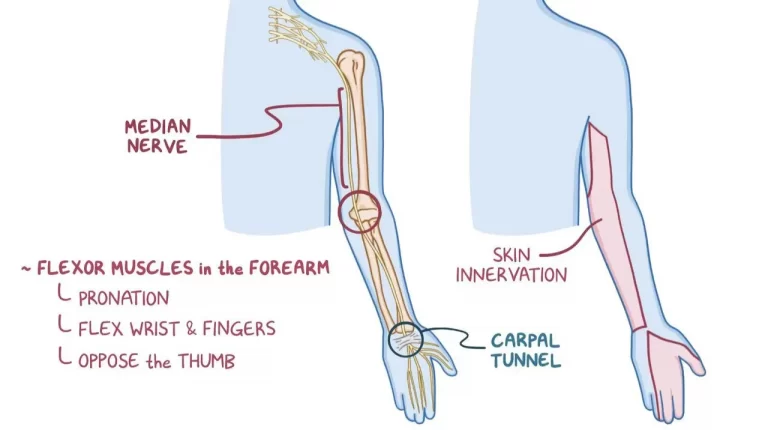
The median nerve (C8-T1) supplies the 1st and 2nd lumbricals. The ulnar nerve (C8-T1) supplies the 3rd and 4th. A fast and effortless way to memorize the lumbrical innervation is given below.
1 2 me, 3 4 you (One two me, three four you)
- 1st and 2nd lumbricals – median nerve
- 3rd and 4th lumbricals – ulnar nerve
Blood supply
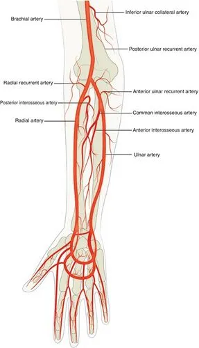
- The majority of the lumbricals’ arteries supply the blood by the dorsal carpal arch, an anastomotic network on the hand’s dorsal surface.
- The 1st and 2nd lumbrical arteries, as well as the 1st and 2nd dorsal metacarpal arteries and the dorsal digital arteries, supply the 1st and 2nd lumbricals.
- The third and fourth lumbrical arteries, as well as the second and third common palmar digital arteries, are branches of the superficial palmar arch, supplying the third and fourth lumbricals.
Functions of lumbricals
- The proximal and distal interphalangeal joints are extended, whereas the metacarpophalangeal joints are flexed.
- The opposing actions are brought about by the way the ligaments cross the MCP on the palmar side but enter distally on the dorsal side of the finger.
- When holding a pen, for example, these coordinated actions are part of the hand’s complex movement and contribute to its total skill.
- Furthermore, the lumbrical muscles of the hand have a long fiber length and a high number of muscle spindles, indicating that they are immersed in proprioception.
Clinical relevance
- Crush injuries to the hand may occasionally result in lumbrical muscle injury.
- Following these injuries, adhesions between the lumbrical and interosseous muscles might develop.
- And the lumbrical muscle goes volar to the ligament while the interosseus muscle passes dorsally, adhesions distal to the inter-palmar plate ligament restrict proximal mobility of the interosseus and lumbrical muscles.
- Because of this malformation, the patient gets intermetatarsal pain when making a fist.
- It is usually treated by relaxing the person’s muscles.
- Carpal tunnel syndrome is also linked to the lumbrical muscles, according to research.
Lumbrical-plus finger
- When the flexor digitorum profundus tendons are separated distally from the origin of the lumbricals, an odd sensation occurs: when trying to close the clenched hand, the fingers stretch out uncomfortably.
- Because of the dissociation of the lower tendons, the lumbricals are momentarily the new attachment location for the flexor digitorum profundus.
- This shows that the individual is moving the lumbricals rather than the flexors.
- Furthermore, because the lumbricals and the flexor digitorum profundus have difficulty in the proximal and distal interphalangeal (PIP) joints, the expected clenched hand finding supports the fingers to expand.
- The lumbrical-plus finger, as it is clinically named, can occur as a result of amputations or accidents.
Lumbrical muscle stretching
- Bend the fingers forward to form an L shape with the hand and fingers while maintaining extended knuckles.
- Repeat using the other hand after holding for a few seconds. 3 times, complete the stretch.
Lumbrical muscle strengthening exercise
Isometric for lumbricals

- Start with sitting in a chair, sit up straight.
- Try to grasp the edge of the table with the knuckles bent and the middle and end joints of the fingers straight.
- Squeeze the edge of the table with the thumb and fingers without bending the fingers’ middle and end joints.
FAQ
The lumbricals are the first and foremost contributors to flexion at the metacarpophalangeal joints as well as an extension at the DIP and PIP joints; although, the interossei also play a minor role in these movements.
Innervation. Lumbrical muscles get nerve supply from the two terminal branches of the tibial nerve; the medial plantar nerve (S1, S2) supplies the first lumbrical muscle. The lateral plantar nerve (S2, S3) supplies the lateral 3 lumbricals.
In palm, lumbricals arise from the sides of tendons of flexor digitorum profundus. It is a hybrid or composite muscle as it is innervated by 2 various nerves.
Flexor Carpi Ulnaris
This large muscle is built for power, flexing and deviating the wrist away from the thumb.