Elbow Plica Impingement Test
The Elbow Plica Impingement Test, also known as the Elbow Flexion Test or the Plica Syndrome Test, is a physical examination technique used to assess for the presence of plica syndrome in the elbow. Plica syndrome refers to the irritation or inflammation of the synovial folds or plicae within a joint, leading to pain and discomfort.
Table of Contents
Introduction
- A common anatomic finding, a synovial plica (fold), occurs in 86-100% of instances; however, symptomatic plica is far less prevalent (7.2-8.7% of all elbow arthroscopies). Synovial plica syndrome is an elbow pain disease caused by symptomatic synovial plica.
- Clinical examination (lateral elbow discomfort) is used to identify synovial plica syndrome, which is often accompanied by local soreness, pain with terminal extension, and/or painful snapping.
- Other elbow disorders, such as tennis elbow, loose bodies, and degenerative arthritis, may look like synovial plica syndrome.
- Although magnetic resonance imaging or ultrasound scans can help in diagnosis when combined with clinical signs, symptomatic plica may also be found as an unexpected discovery during elbow arthroscopy.
- If conservative therapy fails, arthroscopic resection is a viable and safe option.
What is the synovial fold?
- The elbow plica is a prominent fold of the synovial membrane. There have been several variations in location, form, structure, and size recorded. In general, the elbow plica is thought to be physiological. Isogai et colleagues hypothesized that adult folds develop from embryonic structures and undergo major changes from homogeneous structures mixed with the annular ligament to a more heterogeneous appearance in adults. A plica can be used as a basic space filler in non-articular indentations.
- However, it may act as a tension disperser and offer cushioning during the flexion and extension processes. Forces around the elbow gradually transform the synovium into a soft and villous degenerative fold. In rare cases, impinging creates larger, thicker, and harder structures, resulting in mechanical symptoms (pain, cracking, contracture).
The anterior-thin section of the radio-humeral synovial fold occurs in 67-100% of cases.
- Lateral – in 5-20%, a narrow, tiny, crescent, or meniscoid form.
- Posterior – between the larger and lesser sigmoid cavities and radio-humeral surfaces, merges with anterior lateral fold and lateral olecranon fold in 86-100% of cases.
- Lateral olecranon – in 28-33% of cases, on the lateral edge of the olecranon under the anconaeus muscle.
- Circumferential – the continuous plate, integrating anterior and posterior, contributes to 2-12% of the total.
What is elbow synovial plica?
- Synovial plica is an inflammation of a tiny portion of the soft tissues of the elbow joint. To truly understand plica syndrome, you must first grasp the basic anatomy of the elbow.
- The synovial membrane, also known as the synovium, is a connective tissue layer that lines the inside of the joint capsule. It generates synovial fluid, which lubricates the joint and reduces friction between bones during movement.
- During fetal development, this tissue folds inward but is normally resorbed, leaving a tiny empty region in the elbow cavity. However, tissue resorption does not always occur completely. This implies that the typical cavitation of the elbow stays incomplete in many people.
- In other terms, they are born with ‘plicae’.
- Due to the intrinsic suppleness of the synovial membrane, plicae typically interfere with proper joint function.
- However, some situations, such as sitting on a hard surface or using the joint extensively for repetitive movements, can cause them to become irritated and inflamed. When this happens, the joint becomes uncomfortable, and arm mobility may be limited. This condition is known as synovial plica syndrome.
- Synovial plicae are found in more than 80% of the general adult population, according to a medical study. Symptomatic synovial folds, on the other hand, are substantially less prevalent, accounting for fewer than 8% of all elbow arthroscopies.
- Synovial plica can be difficult to diagnose since its symptoms are similar to those of a variety of common elbow disorders.
- However, if a plica is diagnosed as the source of joint discomfort, it is possible to heal successfully and rapidly.
- Elbow synovial plica is an uncommon upper extremity ailment that is sometimes mistaken for Tennis Elbow (another severe elbow condition).
It is generally identified by:
- Sharp or shooting pain in the elbow
- Instability of the elbow joint
- Catching or locking sensation of the elbow.
Other names of elbow synovial plica are:
- Elbow synovial fold syndrome
- Snapping elbow syndrome
- Skipping elbow syndrome.
Synovial plica is more common in athletes and those who participate in repetitive motion activities on a regular basis. For example, hammering or typing.
It can also occur as a result of a direct impact or blow to the elbow.
Symptoms of the elbow synovial fold syndrome
- Dull, chronic arm ache that worsens with elbow usage
- An acute, stabbing, or shooting pain while moving the elbow in a certain posture
- Touch sensitivity on the inner or outer side of the elbow
- Swelling around the elbow
- Reduced range of motion/movement of the elbow
- Increased elbow joint laxity – the sensation that your elbow will ‘give out’ if you try to bend it to a certain degree.
- When bending or straightening your arm, you may feel a snapping, catching, or locking feeling.
- Elbow tenseness
- When you bend or stretch your elbow, you may hear a clicking or ‘popping’ sound.
Purpose
The Elbow Plica Impingement Test is used to diagnose elbow plica syndrome or synovial fold syndrome.
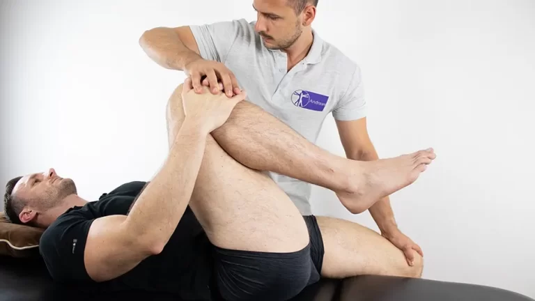
Technique

Posterolateral radiocapitellar plica examination
Step 1: The patient is in a standing or sitting position.
Step 2: Extend the patient’s elbow and pronate the forearm.
Step 3: The examiner applies manual force to the posterolateral portion of the radiocapitellar joint while passively flexing the elbow.
Step 4: Tenderness at the radiocapitellar joint at low flexion angles that decreases significantly at more than 90 degrees of flexion with manual compression force maintained is a positive test.
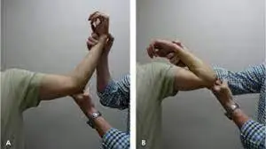
Flexion-Pronation plica test
Step 1: The patient is in a standing or sitting position.
Step 2: The patient’s elbow is stretched and his or her forearm is pronated.
Step 3: While passively flexing the elbow, the examiner provides a lateral force on it.
Step 4: A positive test is indicated by snapping above 90 to 110 degrees of flexion.
Evidence
The Posterolateral radiocapitellar plica test was performed on 24 individuals, with 20 of them testing positive.
- Statistics are explained on the test diagnostics page.
- This test has an 83.3% sensitivity.
- This test has a specificity of 87.5%.
- Positive predictive value -74.1%
- Negative predictive value – 92.5%
- The accuracy value is 86.3%.
Only 14 individuals were studied for the Flexion-Pronation plica test, and only half of them (50%) proved positive.
FAQ
Clinical examination (lateral elbow discomfort) is used to identify synovial plica syndrome, which is often accompanied by local soreness, pain with terminal extension, and/or painful snapping. Other elbow disorders, such as tennis elbow, loose bodies, and degenerative arthritis, can mirror synovial plica syndrome.
In conclusion, painful cracking and popping on the outside of the elbow is frequently caused by inflamed Plicas, fortunately, are usually efficiently treated by excision with elbow arthroscopy.
Plica syndrome is often treated with physical therapy to increase mobility and strengthening to reduce strain and discomfort. Physical therapists are specialists in movement. They improve people’s lives by providing hands-on treatment, patient education, and prescribed mobility.
Ice: Using ice packs on a regular basis helps to relieve pain and inflammation. Anti-Inflammatory Medication: Nonsteroidal anti-inflammatory drugs (NSAIDs) such as ibuprofen can also help decrease pain and inflammation.