
Dinner fork deformity
Table of Contents
Definition
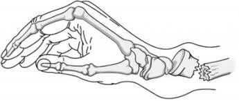
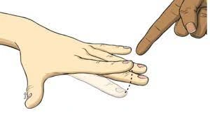
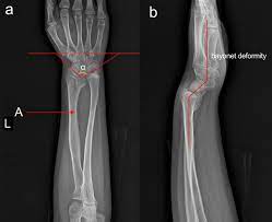
A dinner fork deformity also identifies as a bayonet deformity, happens as the result of a malunited distal radial fracture, generally a Colles fracture. The distal fragment is dorsally angulated, displaced, and often also impacted.
What are the Causes of Dinner fork deformity?
- Wrist fracture mainly after colle’s fracture
- Fall on Overstretched hand (Most common in children)
- People who are suffering from osteoporosis
- Traumatic accident
- Sportsmen, skiers .skaters, and bikers.
- Calcium deficiency is not the exact cause but a contributing factor for the deformity.
Symptoms in Dinner fork deformity
- The person finds difficulty in moving the wrist joint.
- The pain improves when the wrist is flexed.
- There is swelling nearby the wrist area.
- The nearby area becomes tender to the touch.
- Bruising is usual as a result of severe impact.
- There is numbness in the hand.
- Fingers can become pale.
- The person has some difficulty gripping anything.
Findings
- Dorsal tilt
- Radial shortening
- Loss of ulnar inclination
- Radial angulation of the wrist
- Dorsal displacement of the distal fragment.
Diagnosis
- Diagnosis is most frequently made upon the interpretation of posteroanterior and lateral views alone.
Treatment of Dinner fork deformity
Medical treatment
- Symptomatic Medical treatment and It also includes upper limb Elevation, Compression, and Medication.
Surgical treatment
- It incorporates the management based on the severity of the fracture.
- An undisplaced fracture can be treated by only using a cast.
- A fracture with mild angulation and displacement can need closed reduction.
- Significant angulation and deformity can require an open reduction and internal fixation or external fixation.
Physiotherapy Treatment:
Physiotherapy treatment is depends upon the condition of the patient. Mostly after removal of the Cast, Wrist mobilization, stretching of tight muscles around the wrist, deformity corrective splint/brace with exercise and if wrist pain, stiffness is present use of Paraffin wax bath, Transcutaneous electrical nerve stimulation (TENS).
Exercise for Dinner fork deformity
In the first week–
After removal of the cast-
- Look out for the mortar cast any tragedy or an excessive amount of tightness happen or not.
- The sling should be looked out it will be with proper neck cushioning
- Depending on the edema should be treated with height and back rub from fingertip to palm.
- Dynamic Range of movement practice in non-involved side digit, thumb, elbow, and bear joint to forestall solidness.
- On the involved side, just supination and pronation are not permitted generally all other development empower.
In the second week
Recasting should be advised if the cast is too loose or cracked. Start some range of motion exercises.
Wrist extension and flexion

- Rest the forearm on a table.
- Have the hand hang over the ledge with the palm facing down.
- Gently move the hand upward until you feel a slight stretch.
- Lower the hand again to the initial position.
Supination and pronation exercise
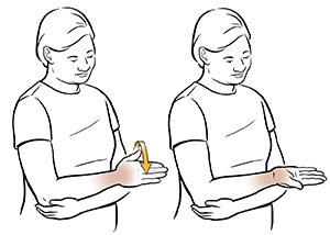
- While seated, lay the hand flat on the leg with the palm facing down.
- Slowly turn the handover, so that the palm is facing up.
Opening and closing fist exercise

- Start with the fist closed (not too tightly).
- Gently open the palm, stretching the fingers outwards.
- Close the fist again.
After the second week –
- Start the wrist mobilization, stretches, and ROM.
Palm facing inward and outward stretch
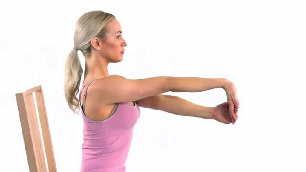
- Start with the arm outstretched in front of you with the palm facing in towards the body and the fingers pointing down towards the floor.
- With the free hand, hold the fingers of the outstretched hand and slowly pull fingers towards you until you experience a slight stretch in the back of the wrist.
- Perform this exercise again, but have the fingers facing UP towards the ceiling with the palm still facing toward the body.
Wrist stretch using a table
- Wrist stretches may also be done by using a table.
- Put both of the palms face down on the edge of a table.
- Fingers point away from you.
- Keep up the elbows straight, and slowly lean the body weight slightly forward.
Palm press (thumbs facing in against body)

- While standing put the hands together in a prayer position in front of the body.
- The elbows will be wide to the sides.
- Draw the shoulders back gently to open the chest.
- Raise the hands gently until they are at face level and bring the elbows and forearms together.
- See if you may keep the palms fully joined, specifically the heels of the hands, and gently lower the hands until you start to feel a gentle stretch.
Palm press (thumbs out)

- While standing, bring the hands together in a prayer position in front of the chest.
- Rotate the fingers down toward the floor. (thumbs now face outward).
- The elbows will be wide to the sides.
- Relax the shoulders down away from the ears.
- Raise the hands gently up, keeping palms pressing together.
Wrist Mobilization
- To lessen torment, edema, and distress – Hydrotherapy and Thermotherapy are given.
Dynamic wrist preparation is started. The patient is made to sit on a seat and keep his lower arm in mid-inclined over a table. With the influenced lower arm settled by the other hand understanding is told to effectively flex and broaden the wrist with gravity disposed of.
The prayer position

- This swift and easy stretch helps with hand and wrist flexibility.
- Place the palms together in a prayer position and leave the elbows on a solid surface like a tabletop.
- Keeping the palms together, lower the sides of the hands until the patient experience a stretch.
- Keep in this position for 5 to 10 seconds and then relax.
- Repeat at least three times.
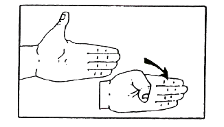
Hand/finger tendon glide
- Initiate with the fingers extended straight out, make a hook fist (fold the first two digits of the fingers towards the palm) and
- then straighten the fingers.
- Next, take a full fist and straighten the fingers again.
- Finally, make a straight fist (keeping the fingertips extended, fold-down until tips touch the lower palm) and then release.
Thumb flexion and extension
- This one is easy.
- Simply hold the thumb up like a person is giving the “OK” sign, and then move the thumb towards the palm of the hand.
- Repeat this for added dexterity.
- Squeeze.
- This exercise is best for mobility problems.
Finger lifts
- Place the hand’s palm down on a flat surface.
- Starting with the thumbs, lift each gently followed by each finger.
- Repeat 8 to 10 times to improve dexterity.
Squeeze a tennis ball
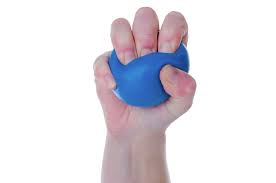
- A tennis ball is the better way to exercise the hand.
- It permits a patient to get the fingers limbered up while improving overall hand strength.
- Begin by squeezing a tennis ball for some time and then releasing it.
- Try this a few times.
- After a few reps, The person will notice that it helps to build up the hand muscles if you keep at it each week.
C” and “O” exercises
- Begin with a straightened hand, pretend you are catching a ball by making a “C” or “O” shape with the fingers, going as far as a person can.
- Straighten the fingers and do it again a few times on each hand to help with flexing issues.
FAQ
The distal fragment is dorsally angulated, displaced, and frequently also impacted. The term is descriptive, as the lateral view of
the wrist is equal to the shape of a fork, seen from the side, tines down.
The Colles fracture is diagnosed as a distal radius fracture with dorsal comminution, dorsal angulation, dorsal displacement,
radial shortening, and a connected ulnar styloid fracture. The term Colles fracture is frequently used eponymously for distal
fractures with dorsal angulation.
Colles Fracture – This is the most usual type of fracture and results in a gross change in the appearance of the wrist, frequently
known as “dinner fork” deformity, where the injured bone angles upward toward the back of the hand. The median nerve near the
wrist can also be injured causing carpal tunnel syndrome.
A dinner fork deformity also identifies as a bayonet deformity, happens as the result of a malunited distal radial fracture, usually a
Colles fracture. The distal fragment is dorsally angulated, displaced, and often also impacted.
A distal radius fracture is an injured around the wrist (distal) end of the radius bone, where it is particularly vulnerable.

One Comment