
Axillary Artery
Table of Contents
Introduction
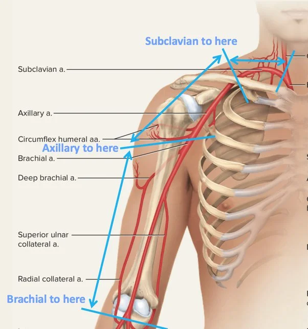
The axillary artery is a crucial blood vessel located in the upper limb, playing a vital role in the vascular supply to the arm, shoulder, and thorax. It is a direct continuation of the subclavian artery and extends from the outer border of the first rib to the lower border of the teres major muscle, where it becomes the brachial artery.
The upper limb, axilla, or what is popularly called the armpit, and thorax are some of these bodily components. The human body’s thorax extends from the neck to the abdomen.
The arm extending from the shoulder to the hand is considered an upper limb. The subclavian artery starts where it crosses the first rib and continues as the axillary artery. It becomes the brachial artery as it descends. One can separate the axillary artery into three pieces. The position of these components in relation to the pectoralis minor muscle, which is situated in front of the axillary artery, serves as a distinguishing factor.
The first segment of the artery is located medially, or close to the middle, of the pectoralis minor muscle. The second segment of the artery is directly behind the pectoralis minor muscle. The third portion of the lateral artery (beyond or further from the middle) connects to the pectoralis minor muscle.
The thoracic, upper limb, and axillary regions receive oxygenated blood through the cooperative efforts of the three components of the axillary artery. Blood is returned to the heart by the axillary vein, which runs parallel to the axillary artery.
What is an axillary artery?
The axillary artery is a sizable muscle vessel that passes through the axilla. It is in charge of delivering blood rich in oxygen to the upper limb and the musculocutaneous system, including the upper lateral thorax and scapula.
As the subclavian continuation, the axillary artery begins at the outside edge of the first rib and finishes at the lower border of the Teres major tendon, when it acquires the name brachial.
The direction of the vessel changes depending on the limb’s location. For example, when the arm is angled at a right angle to the trunk, the vessel is almost straight; when the arm is raised above the trunk, it concave upward; and when the arm is by the side, it convex upward and laterally. The artery is quite deeply positioned at its origin, however, it is superficial toward its termination, covered only by fascia and skin.
Structure:
A common description of the axillary artery is that it is divided into three sections according to where it lies about the superficial pectoralis minor muscle.
First section: The portion of the artery superior to the pectoralis minor
Second section: The portion of the artery posterior to the pectoralis minor
Third section: The section of the artery inferior to the pectoralis minor
Course:
It passes anterior to the teres major muscle and posterior to the pectoralis minor as it descends in the axilla posterior behind the axillary vein. At the inferior boundary of the teres major, it continues as the brachial artery.
Where the subclavian artery ends at the outer edge of the first rib, it continues as the axillary artery.”
The brachial plexus’s lateral (superiorly), posterior (posteriorly), medial (inferiorly), and anteriorly bordered ansa pectoralis cords surround it as it subsequently passes into the axilla.
The pectoralis minor muscle travels superolaterally from its origin at the third, fourth, and fifth ribs to its insertion on the medial portion of the coracoid process, dividing the artery into three parts as it passes through the axilla. Each segment conveniently gives the same amount of branches. Three branches depart from the third section below, two from the second segment below, and one from the first segment above (all relative to pectoralis minor). The axillary artery becomes the brachial artery as it leaves the axilla and continues into the arm at the lower edge of the teres major.
Axillary artery in the axilla. The axillary artery becomes the brachial artery as soon as it leaves the axilla.
Sections:
The relationship between the axillary artery and the pectoralis minor muscle divides it into three sections. It is split into three sections to make the description of the vessel easier to read: the first section is located above the Pectoralis minor, the second part is behind it, and the third part is below the pectoralis minor:
First (suprapectoral) portion: from the superior border of the pectoralis minor to its starting point at the lateral edge of the first rib
The lateral anterior thoracic nerve, the thoracoacromial and cephalic veins, the clavicular portion of the Pectoralis major, and the coracoclavicular fascia cover the anterior portion of the axillary artery; posterior to it are the first intercostal space, the corresponding Intercostalis externus, and the brachial plexus, which is divided from it by a small area of areolar tissue on its lateral side; on its medial, or thoracic, side is the axillary vein, which crosses over the artery; the first and second digitations of the Serratus anterior; the long thoracic and medial anterior thoracic nerves; and the brachial plexus’s medial cord. It is encased in a fibrous sheath called the axillary sheath, which is continuous with the deep cervical fascia above, along with the brachial plexus and the axillary vein.
Second (retropectoral) portion: posterior to the pectoralis minor
The Pectorales major and minor cover the second segment of the axillary artery anteriorly; The axillary vein is located on the medial side, separated from the artery by the medial cord of the brachial plexus and the medial anterior thoracic nerve; the lateral side is home to the lateral cord of the brachial plexus. Behind it is the posterior cord of the brachial plexus and some areolar tissue that acts as an intermediary between it and the Subscapularis. Thus, the artery is surrounded by the brachial plexus on three sides, which also keeps it isolated from the vein and surrounding muscles.
Third (infrapectoral) portion: From the inferior border of the pectoralis minor to the inferior border of the teres major
The third segment of the axillary artery runs from the Pectoralis minor’s lower border to the Teres major tendon’s lower border. Its lateral side is the Coracobrachialis, and its medial or thoracic side is the axillary vein. In front, it is covered by the lower portion of the Pectoralis major above, but only by the integument and fascia below; in back, it is related to the lower part of the Subscapularis, as well as the tendons of the Latissimus dorsi and Teres major.
The brachial plexus nerves are related to this section of the artery in the following ways: the lateral head and trunk of the median are on the lateral side, and the musculocutaneous nerve is located a short distance away; The ulnar (between the vein and artery) and medial brachial cutaneous (to the medial side of the vein) are located on the medial side; in front are the medial head of the median and the medial antibrachial cutaneous, the radial and axillary are located behind, the latter only extending to the lower border of the Subscapularis.
Relations:
Along the whole course of the axillary artery, the axillary vein is located medial to it.
The brachial plexus encircles the axillary artery in the axilla. The locational descriptions of the brachial plexus cords are based on the second segment of the axillary artery. For example, the brachial plexus’s posterior cord gets its name from the fact that it is located behind the artery’s second part.
Relationships between the axillary artery’s first segment:
- Anterior: pectoralis major, clavicle, and platysma;
- Posterior: the brachial plexus’s medial chord and the serratus anterior muscle;
- Lateral: the brachial plexus’s posterior cord;
- Medial: axillary vein,
Relationships between the axillary artery’s second segment:
- Anterior: the pectoralis minor muscle
- Posterior: the brachial plexus’s posterior cord;
- Lateral: the brachial plexus’s lateral cord;
- Medial: the axillary vein and the brachial plexus’ medial cord.
Relationships between the axillary artery’s third segment:
- Anterior: the pectoralis major;
- Posterior: main muscles of the teres major and latissimus dorsi;
- Lateral: coracobrachialis muscle
- Medial: axillary vein
Branches:
The axillary artery has several smaller branches. These branches have very different origins; for example, the anterior and posterior circumflex arteries may share a trunk. The name of an arterial branch refers to its path rather than where it originates.
The branches are:
- First part (1 branch)
- Superior thoracic artery (Supreme thoracic artery)
- Second part (2 branches)
- Thoraco-acromial artery
- Lateral thoracic artery. The lateral thoracic artery will most likely branch from the following (in order of likelihood) if it does not originate from the axillary artery: The thoracoacromial, suprascapular, subscapular, and third portion of the axillary arteries are the first four.
- Third part (3 branches)
- Subscapular artery
- Anterior humeral circumflex artery
- Posterior humeral circumflex artery
The axillary artery shows considerable variability, with just 10% of people having the traditional configuration of six distinct branches. The subscapular and posterior circumflex humeral arteries arise from a similar trunk origin in around 20% of people. The anterior and posterior circumflex humeral arteries arise from the same trunk of origin in a comparable percentage. Instead of coming from the brachial artery, the deep brachial artery originates from the axillary artery in 10–15% of cases or the posterior circumflex humeral artery in 5% of cases.
- First section
The superior thoracic artery supplies the pectoralis major and minor by running anteriorly.
Superior thoracic artery:
The axillary artery’s first branch is the superior (highest) thoracic artery. It is released near the anterior scalene muscle’s outer border. It is a component of the arterial supply to the pectoral muscle.
- Second section
The thoracoacromial artery supplies the clavicle, deltoid, acromion, and pectoralis muscles by its four terminal branches after passing across the pectoralis minor to breach the clavipectoral fascia.
The lateral thoracic artery supplies the serratus anterior and both pectoralis muscles, as well as serving as a vital blood supply to the breast in females. It runs laterally along the bottom border of the pectoralis minor.
Thoracoacromial artery
Two vessels originate from the second segment of the axillary artery. Four additional arteries arise from the thoracoacromial (acromiothoracic) artery, which is a major trunk. The thoracoacromial artery penetrates the clavipectoral fascia by its branches, supplying the indicated upper limb and trunk regions.
- The clavicular branch supplies the sternoclavicular joint and subclavius muscle as it passes superomedially in that direction.
- The pectoral branch supplies oxygenated blood to the muscles and mammaries of the pectoralis by moving inferomedially toward them.
- Deep inside the deltoid muscle, the acromial branch passes through the coracoid process’ medial edge. It supplies this muscle and then penetrates it to join the acromial anastomosis at the acromion.
- Lastly, the deltoid (humeral) branch passes via the deltopectoral groove (where it meets the cephalic vein) and across the tendon over the pectoralis minor. It supplies the deltoid and pectoralis major muscles along this path.
Lateral Thoracic artery
The second branch of the axillary artery is called the lateral thoracic artery.. It moves along the inferior pectoralis minor muscle’s edge inferomedially.
The lateral thoracic artery supplies the thoracic wall’s structural components with oxygenated blood.
- Muscles of the Serratus anterior
- Pectoralis muscle
- Mammaries
- Subscapularis muscle
- Third Part
Subscapular artery, anterior and posterior humeral artery
Subscapular Artery
The major branch of the axillary artery, the subscapular artery, originates from the third section of the artery. Originating at the inferior edge of the subscapularis muscle, the subscapular artery extends distally to the inferior angle of the scapula. After travelling caudally, the subscapular artery splits into the thoracodorsal artery and the circumflex scapular artery.
The circumflex scapular artery enters the infraspinatus fossa by passing through the (upper) triangular gap on the lateral margin of the scapula. It meets the scapular anastomosis at this point. The latissimus dorsi muscle is supplied by the thoracodorsal artery, which continues inferiorly next to the thoracodorsal nerve. The internal intercostal, internal mammary, and pectoral branches of the thoracoacromial arteries create an anastomosis with the thoracodorsal branch of the subscapular artery.
Circumflex humeral arteries
Ultimately, the anterior and posterior circumflex humeral arteries (ACHA & PCHA, respectively) emerge from the third segment of the axillary artery. Between the two arteries, the ACHA is the smaller one. It moves horizontally, deep to the short head of the biceps brachi and coracobrachialis and toward the surgical neck of the humerus. Running along the anterior portion of the humeral surgical neck, the anterior circumflex humeral artery anastomoses with its posterior counterpart It branches at the intertubecular groove, going superiorly via the sulcus to supply the glenohumeral joint.
The PCHA passes via the quadrangular space posterior to the axillary nerve. After that, the PCHA runs anteriorly around the humerus’ surgical neck, supplying the deltoid muscle and shoulder joint while anastomosing with the profunda brachii and ACHA.
Clinical importance
The only place where clamping the axillary artery safely won’t put the arm in risk is close to the subscapular artery’s origin (and far from the subclavian artery’s thyrocervical trunk). The dorsal scapular artery and suprascapular artery are two arteries that can send collateral circulation to the arm via the anastomotic network encircling the scapula.
In cardiac surgery, the right axillary artery is frequently utilized as an arterial cannulation site, especially when replacing the ascending aorta and aortic arch and repairing aortic dissection.
FAQs
Where is the axillary artery located?
At the outer edge of the first rib, the axillary artery splits off from the subclavian artery, and it ends at the outer edge of the teres major muscle, where it becomes the brachial artery.
Why is the axillary artery important?
The main artery supplying the upper limb is the axillary artery, which starts as the subclavian artery and continues into the axilla after emerging from under the first rib.
How to palpate the axillary artery?
Using his or her index and middle fingers, the anesthesiologist palpates the patient’s axillary artery. The anesthesiologist locates the artery and then holds his or her fingers in position to precisely identify the needle insertion spot.
What is the main artery in the arm?
The primary artery of the upper extremity, the brachial artery is the axillary artery’s extension, beginning at the lower edge of the teres major muscle. The brachial artery runs along the arm’s ventral side, giving rise to several minor branching arteries prior to arriving at the cubital fossa.
What vein and nerve are found in the axillary artery?
The medial and lateral pectoral nerves, the medial cord of the brachial plexus, the ulnar nerve, and the medial cutaneous nerve of the arm are among the neurovascular structures of the axilla that are carried in a bundle by the axillary artery and vein.
Reference:
- Posterior circumflex humeral artery. (2018, January 23). Healthline. https://www.healthline.com/human-body-maps/axillary-artery#1
- Micheau, A., & Hoa, D. (2024, March 18). Axillary artery. e-Anatomy – IMAIOS. https://www.imaios.com/en/e-anatomy/anatomical-structure/axillary-artery-1553672960
- Axillary Artery | Complete Anatomy. (n.d.). www.elsevier.com. https://www.elsevier.com/resources/anatomy/cardiovascular-system/arteries/axillary-artery/16785
- Axillary artery. (2023, October 30). Kenhub. https://www.kenhub.com/en/library/anatomy/the-axillary-artery
- Hacking, C., & Jones, J. (2013). Axillary artery. Radiopaedia.org. https://doi.org/10.53347/rid-24582
- Axillary artery. (2024, May 2). Wikipedia. https://en.wikipedia.org/wiki/Axillary_artery#:~:text=In%20human%20anatomy%2C%20the%20axillary,is%20called%20the%20subclavian%20artery.
One Comment