Prone instability test
Table of Contents
Introduction
The Prone Instability Test (PIT) is a clinical assessment used to evaluate the presence of instability in the lumbar spine. It is primarily used by healthcare professionals, such as physical therapists and chiropractors, to assess patients with low back pain.
The Prone Instability test is an orthopedic test used to evaluate radiographic lumbar instability, which is one of the most common causes of chronic low back pain.
Several authors examined this test for inter-rater reliability and found Kappa values ranging from 0.46 (poor) to 0.87 (high).
However, when it came to evaluating radiographic lumbar instability, it revealed low sensitivity and specificity values, making this test a poor diagnostic tool in clinical practice.
Technique of Prone instability test
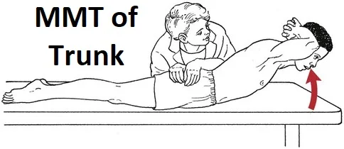
- The examination is divided into two sections. During the first section, you instruct the patient to lie prone with his legs down from the bench but in contact with the ground. Then you apply PA pressure to various parts of the lumbar spine in an attempt to cause discomfort.
- If the patient complains of pain, you advise him to elevate his legs off the ground and apply PA pressure again. If the patient’s discomfort is eased by actively lifting his legs off the ground, this indicates that the patient can actively stabilize his spine, which is a positive sign.
- A positive test also shows that a patient would benefit from a stability exercise program.
Diagnostic Accuracy for Success with a stabilization exercise program:
- This test has a sensitivity of 72%.
- This test has a specificity of 58% +LR:.48, -LR:1.7 (“Preliminary development of a clinical prediction rule for determining which patients with low back pain will respond to a stabilization exercise program” research participants had non-radicular low back pain).
Importance of the Prone instability test
- Poor lumbar vertebral stability can put you at risk for a variety of diseases, including lumbar spondylolisthesis, spinal cord lesions, and lower extremity musculoskeletal abnormalities.
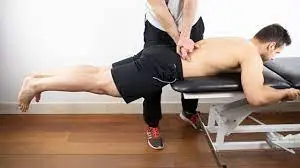
- The off-the-table position participates in the hip and lumbar extensors, which provide to stabilize the hypermobile segment and can help evaluate how much a core stabilization program might benefit the patient. Some books depict the patient extending the legs all the way to the plane of the table, while others show the patient only lifting the legs a few inches off the ground.
- While both methods activate the core muscles, a position with the legs elevated to the plane of the table may provide more stimulation. The weight of the legs has a raised moment arm related to the torso in that elevated position.
- This greater torque exerts more demands on the core muscles, resulting in a stronger contraction. Based on this diagnostic accuracy, it is difficult to draw judgments concerning lumbar instability based simply on this test.
- It is critical to group these results with a + Gower’s Sign, Instability catch, abnormal motion discoveries, and other indicators.
Evidence
- To confirm the indications and symptoms of lumbar instability, this test should be done in combination with other examinations. This test was also included in Hicks’ clinical prediction criteria for individuals who responded well to spinal stabilization exercises.
- As a result, positive test findings were associated with patients who responded favorably to spinal stabilization exercise programs, whereas negative test results were associated with patients who did not respond favorably.
- This test was one of four factors found and reported in CPR for the success and failure of lumbar spinal stabilization exercise programs.
FAQ
If the discomfort is present in the resting position but disappears in the second position, this indicates lumbo-pelvic instability. muscle activation has the ability to stabilize the spinal segment.
Symptoms may include low back pain, discomfort, stiffness, and muscular spasms. Radiculopathy symptoms may include numbness, tingling, pain, or weakness in the legs.