PATELLAR TENDINITIS: Physiotherapy Treatment
Table of Contents
What is patellar tendonitis?
Patellar Tendinitis (Patellar tendinopathy or as it is commonly known patellar tendonitis or tendinitis) is an overuse injury affecting your knee joint. It is the result of your patella tendon being over-stressed. A common name for it is Jumper’s Knee.
Patellar tendinitis is an injury to the tendon connecting your kneecap (patella) to your shinbone. The patellar tendon works with the muscles at the front of your thigh to extend your knee so that you can kick, run and jump.
ANATOMY OF KNEE JOINT
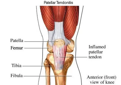
The patellar tendon runs inferiorly from the patella bone to the tibial tuberosity. The patella is a large sesamoid (a bone within a tendon) bone with a triangular transverse cross-section, that lies within the quadriceps tendon.
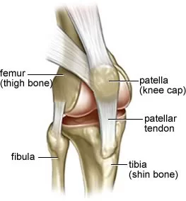
The patellar tendon runs from the apex, adjoining margins, distal posterior surface, and rough anterior surface of the patella to the tibial tuberosity, which is a small bony bump on the anterior aspect of the tibia.
A tendon is a connective tissue that connects a muscle to a bone, and the patellar ‘tendon’ in fact connects a bone to a bone (patella to tibial tuberosity). The correct name is therefore the patellar ligament. The patellar ligament is approximately 5 cm in length. However, its length is not constant and mostly increases from full extension to 30 degrees of knee flexion.
The medial and lateral parts of the quadriceps femoris descend on either side of the patella and are inserted onto the upper anterior surface of the tibia. They merge into a continuous capsule and form the medial and lateral patellar retinacula.
The posterior aspect of the patellar ligament is separated from the knee joint by an infrapatellar fat pad and a synovial membrane. An infrapatellar bursa also separates the patellar ligament from the tibia.
Patellar tendinitis symptoms
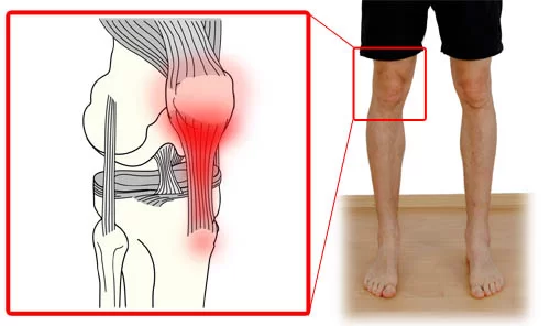
- Knee Pain and tenderness around your patellar tendon
- Swelling around the knee joint
- Knee Pain with jumping, running, or walking
- Pain when bending or straightening your leg
- Tenderness behind the lower part of your kneecap
CAUSES
Patellar tendonitis comes from repetitive stress on the knee joint, most often from overuse in sports or exercise.
The repetitive stress on the knee creates tiny tears in the tendon that, over time, inflame and weaken the tendon.
following factors may cause patellar tendonitis:
- tight leg muscles
- uneven leg muscle strength
- misaligned feet, ankles, and legs
- obesity
- shoes without enough padding
- hard playing surfaces
- chronic diseases that weaken the tendon
Athletes are more at risk because running, jumping, and squatting put more force on the patellar tendon. For example, running can put a force of up to five times your body weight on your knees.
DIAGNOSIS
X-rays: X-rays help to exclude other bone problems that can cause knee pain.
Ultrasound: This test uses sound waves to create an image of your knee, revealing tears in your patellar tendon.
Patellar tendinitis MRI (Magnetic resonance imaging): MRI uses a magnetic field and radio waves to create detailed images that can reveal subtle changes in the patellar tendon.
Patellar tendinitis Treatment
NSAIDs – over-the-counter (OTC) drugs for short-term pain and inflammation reduction.
include:
- ibuprofen (Advil)
- naproxen sodium (Aleve)
- acetaminophen (Tylenol)
If your pain is severe, your doctor may give you a corticosteroid injection in the area around your patellar tendon. This is more effective in reducing severe pain. However, the Mayo Clinic reports that it also may weaken the tendon and possibly make it more likely to rupture. So, it’s important to put considerable thought into this treatment and its associated risks.
Corticosteroid injection: An ultrasound-guided corticosteroid injection into the sheath around the patellar tendon may help relieve pain. But these types of drugs can also weaken tendons and make them more likely to rupture.
Platelet-rich plasma injection: This type of injection has been tried in some people with chronic patellar tendon problems. Studies are ongoing. It is hoped the injections might promote new tissue formation and help heal tendon damage.
Oscillating needle procedure: This outpatient procedure is performed using local anesthesia. Your doctor uses ultrasound imaging to guide a small oscillating needle that cuts away the damaged area while sparing a healthy tendon. This is a relatively new procedure, but results have shown promise.
Patellar tendonitis surgery:
Surgery: In rare cases, if other treatments fail, your doctor might suggest surgical debridement of the patellar tendon. Some procedures can be done through small incisions around your knee.
Patellar tendinitis Physical therapy:
Physical therapy are pain relieving electrotherapy modalities and exercise.
- Ice Pack / Hot Pack: Apply ice or Hot Pack for 20 to 30 minutes, 4 to 6 times per day, especially after activity.
- Ultrasound Therapy or phonophoresis (ultrasound-delivered medication) may decrease pain symptoms.
- Interferential Therapy (IFT)
- TENS (Transcutaneous Electrical nervous stimulation)
Patellar tendinitis exercises:
The following exercise is helpful.
1. Isometric Quad Contraction Exercise:
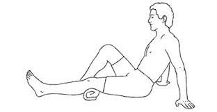
Lie on your back with the leg you want to exercise straight. Place a small rolled towel underneath the knee. Slowly tighten the muscle on top of the thigh (quadriceps) and push the back of the knee down into the rolled towel.
Hold the contraction for 5 seconds and then slowly release, resting 5 seconds between each contraction. Perform 3 sets of 10 repetitions, 1 time daily.
2. SITTING QUADRICEPS EXERCISE
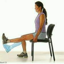
Sit on a chair with both legs bent at 90°.
Slowly raise your right leg so that it’s parallel to the floor, keeping your left foot on the ground.
Hold for 30 seconds, then slowly bring the right foot back to the floor, and repeat on the left leg.
Do 10 times twice a day.
3. Straight leg raise exercise (SLR):
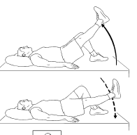
Lie on your back with your legs straight out in front of you. Bend the knee on your uninjured side and place the foot flat on the floor. Tighten the thigh muscle on your injured side and lift your leg about 8 inches off the floor. Keep your leg straight and your thigh muscles tight. Slowly lower your leg back down to the floor. Do 2 sets of 15.
4. SIDE LEG RAISES:
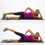
Lie on one side with your legs stacked. Bend the bottom leg for support. Straighten the top leg and raise it to 45 degrees. Hold for 5 seconds, lower and relax briefly, then repeat 10-15 times. Switch sides and start over. Want to try a bit of a different spin on the move? Point the toe of your upper leg slightly toward the floor as you raise it.
5. Prone hip extension exercise:

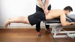
Lie on your stomach with your legs straight out behind you. Fold your arms under your head and rest your head on your arms. Draw your belly button in towards your spine and tighten your abdominal muscles. Tighten the buttocks and thigh muscles of the leg on your injured side and lift the leg off the floor about 8 inches. Keep your leg straight. Hold for 5 seconds. Then lower your leg and relax. Do 2 sets of 15.
6. Squats exercise:
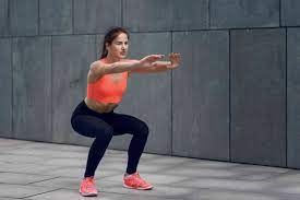
Stand with your feet shoulder-distance apart and stretch your arms out in front of you.
Slowly bend your knees until you’re in a half-sitting position. Hold on to a chair for balance, if necessary.
Keep your back straight and chest lifted — don’t lean forward.
With your feet flat on the floor, hold the position for 5 seconds, then slowly stand back up.
There should be no pain while performing this exercise. Do 10 repetitions, and slowly work up to three sets of 10.
7. Lunges:
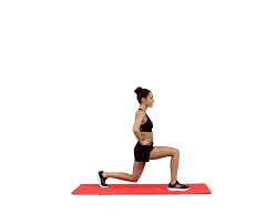
Take a big step forward with your right leg and start to shift weight forward so your heel hits the floor first.
Lower body until right thigh is parallel to the floor and right shin is vertical (it’s okay if knee shifts forward a little as long as it doesn’t go past right toe). If mobility allows, lightly tap the left knee to the ground while keeping weight on the right heel.
Press into the right heel to drive back up to the starting position.
Repeat on the other side.
8. Rectus Femoris muscle Stretching exercise:
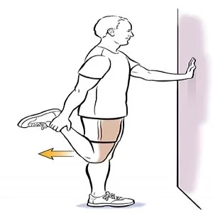
Kneel on your injured knee on a padded surface. Place your other leg in front of you with your foot flat on the floor. Keep your head and chest facing forward and upright and grab the ankle behind you. Gently bring your ankle back toward your buttocks until you feel a stretch in the front of your thigh. Hold for 15 to 30 seconds. Repeat 2 to 3 times.
9. Hamstring Stretching exercise:
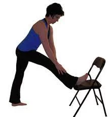
Put the heel of the leg on your injured side on a stool about 15 inches high. Keep your leg straight. Lean forward, bending at the hips, until you feel a mild stretch in the back of your thigh. Make sure you don’t roll your shoulders or bend at the waist when doing this or you will stretch your lower back instead of your leg. Hold the stretch for 15 to 30 seconds. Repeat 3 times.
10. CALF STRETCH:
Stand facing a wall with the leg to be stretched behind you and the other leg in front. Place your hands or forearms on the wall for support. Slowly bend the front knee, keeping the heel of the leg behind you down on the floor. Once you feel a stretch in your calf muscle at the back of your ankle, hold for 30 seconds. Slowly relax. Perform 3 repetitions
11. Prone knee bends exercise
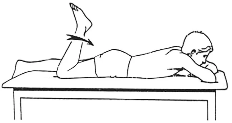
Lie on your stomach and slowly bend your injured knee up, using your hamstring on the back of your thigh.
DO’S & DON’TS
Do exercise daily: Regular cardio exercise strengthens the muscles and increases the flexibility of your knee. It includes walking, swimming, water aerobics, and stationary cycling.
Do use the “RICE” Principle: Rest, ice, compression, and elevation are beneficial for Knee pain which is caused by minor injury. Give rest to your Knee then apply ice to reduce swelling, and wear a bandage to keep your knee elevated.
Buy a patellar tendon strap. This strap is a band that goes around your leg just under the patella. The strap puts pressure on the tendon, thereby redistributing the load throughout the tendon and reducing pain
Don’t play through pain. As soon as you notice exercise-related knee pain, ice the area and rest. Until your knee is pain-free, avoid activities that put stress on your patellar tendon



2 Comments