Cardiac embolism
Table of Contents
What is a cardiac source of embolism?
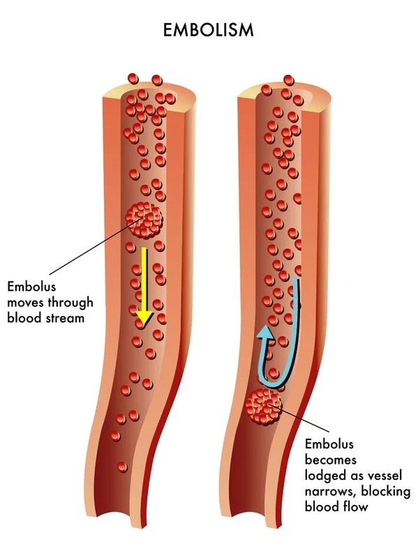
A cardiac embolism is an obstruction that travels from the heart to a section in a blood vessel. An embolus can be prepared of fatty material, or it container be a blood clot. Many factors can provide the formation of blood clots. A cardiac root of embolism can travel to a different position in the body, possibly resulting in life-threatening complications.
Blood vessels can become blocked by a progressive increase of material in the vessel wall. This is how atherosclerosis creates blockages. When plaque or another substance, similar to a blood clot, travels through the bloodstream and becomes squees in a blood vessel because the diameter of the vessel is onward small for it to progress, it is called an embolism. A cardiac embolism is an inhibition that travels from the heart to a section in a blood vessel. An embolus can be prepared of fatty material, or it container be a blood clot.
What is Cardiac embolism?
A cardiac embolism sources a blocked artery, which can affect blood flow. They are caused by a cardiac embolus, which flows in the heart. A cardiac thrombus is a blood clot that conformation in the heart, and when it travels, it is called an embolism.
Depending on where the blockage takes place, a person can experience different complications, for example:
- A blockage in the heart’s blood vessels container leads to a heart attack.
- A blockage in the organs container leads to organ damage.
- A blockage in the limb container leads to a peripheral embolism.
- When a cardiac embolus makes its approach into the brain, it can cause a cardiac embolism, which container cause what doctors call a cardioembolic stroke. These strokes make up around 25%Trusted Source of all ischemic strokes
Symptoms of Cardiac embolism
shortness of breath
soreness in the arms, neck, back, or jaw
chest pain
A person may also experience:
sweating
lightheadedness
nausea
Causes of Cardiac embolism
a blood clot
air
fat
tumor
- atrial fibrillation: atrial fibrillation is the common cause of cardioembolic stroke because the blood clots from the left atrial accessory in the heart travel to the brain.
- heart failure: this weakens the heart. blood clots container form, as the heart is impotent to pump blood around the body.
- Atherosclerosis: Atherosclerosis is the increase of fats and cholesterol in the artery walls. Plaque can increase and restrict the blood flow in the arteries. If the plaque ruptures, the fats and cholesterol container travel through the body, leading to a blood clot.
- Vasculitis: Vasculitis causes the blood vessels to enhance inflamed. Clots container form when the platelets stick to the damaged blood vessels.
Risk factors
The following Risk factors may vary depending on the cause of embolus such as:
- having obesity
- smoking
- taking oral contraceptives
- being over age 60
- a family history of blood clots
- diabetes
- high blood pressure
- high cholesterol
- having heart failure
- having atrial fibrillation
- An air embolus container occurs as a result of scuba diving and during some medical procedures.
Risk factors for fat embolisms combine:
being young in age
closed bone fractures
multiple bone fractures
long-term conservative treatment for fractures, parallel as casts and splints
Diagnosis
If a cardiac embolism is suspected one or more tests may be required to confirm the diagnosis.
- electrocardiograms, which measure the heart’s activity
- transesophageal or transthoracic echocardiograms, evaluate heart functionality and structure and look for clots in the heart.
- MRI and CT scan to measure brain function or look at blood vessels in the brain, the neck, or around the body
- blood tests to assess for damage to the heart muscle
- carotid ultrasound, which is an imaging test that examines the inward of the carotid arteries.
Treatment of Cardiac embolism
With cardiac embolism, the first goal is to prevent the blood clots from producing in the first place. If you are at risk for blood clots, it medical team may prescribe blood-thinner medications known as anticoagulants. If the risk for blood clots is the result of a specific condition, similar to atrial fibrillation (Afib or AF), then medications may be prescribed to adjust the pace or the rhythm of the heartbeat. If your condition does not respond to these medications or if you are inadequate to tolerate their side effects, medical procedures are available that container reduces the frequency of the Afib episodes, treat the cells that are causing the atrial fibrillation, or prevent blood from connecting in the LAA. These therapies include:
In electrical cardioversion, there the heart is treated with an electric shock to restart its normal sinus rhythm. Read about electrical and pharmacologic cardioversion at the moment.
Catheter ablation is a procedure in which the doctor destroys a small quantity of heart tissue that is sending irregular electrical signals that cause atrial fibrillation. Learn about catheter ablation here.
Left atrial appendage closure (or LAAC), is a minimally invasive procedure that authorization. apart the LAA to prevent blood from collecting, and possibly clotting, in this part of the heart. To understand more about this procedure, visit the Seconds Count Left Atrial Appendage Closure Center.
Physical therapy
We constantly apprehend that physical therapy (PT) helps patients get back on their feet after an injury, surgery, stroke, or another medical issue. Physical therapists (PTs) container also play an important role in identifying signs and symptoms of a potential blood obstruction and in helping patients recover from clots. To learn how PTs can help with blood clot prevention and management, we turned to who specializes in PT for the heart and lungs (also known as cardiopulmonary PT).
If a PT understands the symptoms of a blood clot (such as pain or swelling in the leg or unexplained shortness of breath). These devices allow PTs to input symptoms and other factors, like whether a patient is a smoker if the patient has reduced mobility for more than three days, if they are obese, etc. the PT might refer a patient for additional testing. PTs can directly refer a patient for testing (like an ultrasound); in another state, the patient will be mentioned to a healthcare provider for further evaluation.
How to Prevent Cardiac embolism?
The determent of a cardiac embolism depends on the potential hazard. If a person is at risk of developing blood clots, a healthcare professional may formulate blood-thinning medication, parallel to anticoagulants.
If a person has a heart condition, similar to atrial fibrillation, they may take medications to help regulate their heart’s rhythm.
A healthcare professional may either perform the following procedures.
Electrical cardioversion: An electric shock container restart the heart’s normal rhythm.
Catheter ablation: A doctor destroys the small quantity of heart tissue that is sending signals that cause atrial fibrillation. Left atrial attachment closure: This is a procedure to help prevent blood from collecting and clotting.
If a person is at risk of heart failure, taking steps to prevent this container help reduce the chance of clots. A person container holds the following steps to reduce the risk of heart failure: trying to stop smoking, if applicable eating heart-healthy foods, such as:
- fruit
- vegetables
- low-fat dairy
- lean proteins
- trying to maintain a moderate weight
- monitoring and managing any existing heart conditions
If the embolism consists of fat or air, a healthcare professional may approve the surgery.
FAQs
A cardiac source of embolism causes a blocked artery that occurs as a result of a blood clot that forms in one of the chambers of the heart. Blood clots in the heart container occur due to a variety of reasons, including parallel atrial fibrillation or cardiomyopathy/congestive heart failure.
Coronary embolism is a rare and potentially fatal phenomenon that happens primarily in patients with valvular heart disease and atrial fibrillation.
A blood clot in the heart or lungs could include symptoms such as chest pain, shortness of breath, upper body discomfort in the arms, back, neck, or jaw, recommended heart attack, or pulmonary embolism.
The left atrium and left atrial appendage are the most ordinary sources of cardiac emboli. Thrombus formation is strongly associated with atrial arrhythmias (atrial fibrillation, atrial flutter) and the massive majority of thrombi arise in the left atrial appendage.
Massive pulmonary embolism (PE) without cardiac arrest has to arise associated with a mortality rate of 30%. However, when cardiac arrest follows, mortality may be as high as 95%.
Embolic strokes are usually caused by a blood clot that forms abroad in the body (embolus) and travel through the bloodstream to the brain. Embolic strokes continually result from heart disease or heart surgery and occur rapidly and without any warning signs.
Thrombosis occurs when a thrombus, or blood clot, expands in a blood vessel and reduces the flow of blood through the vessel. An embolism occurs when a piece of a blood clot, foreign object, or other bodily substance enhances stuck in a blood vessel and largely obstructs the flow of blood.
These blood-thinning prescriptions are called anticoagulants to prevent existing clots from getting bigger and new clots from forming while your body works to break up the clots. Heparin is a frequently used anticoagulant that can be given in a vein or injected under the skin.
How to prevent blood clots from stopping smoking – if you smoke, quitting is the best effect you can do on your health.
getting regular exercise. not sitting still for a long time – such as watching TV or using a computer. avoid becoming dehydrated. maintaining a healthy weight, along with eating a healthy balanced diet.


