Accessory Navicular Syndrome
Table of Contents
Introduction
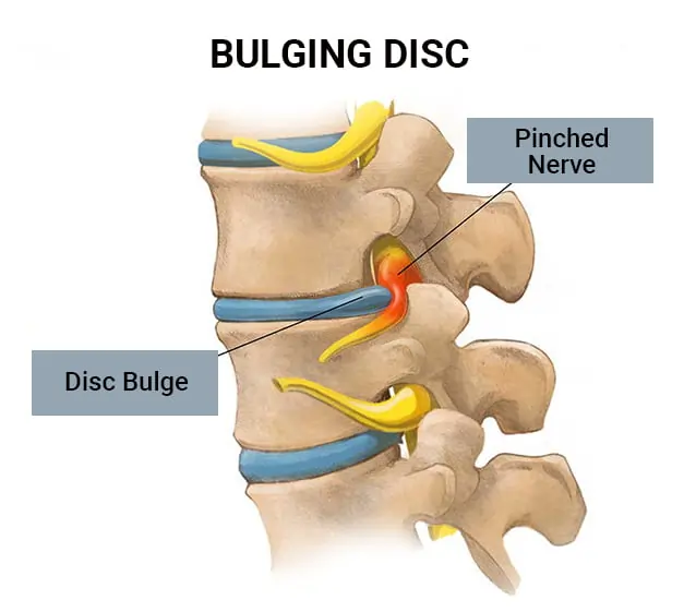
Accessory Navicular Syndrome (ANS) is a relatively common condition that affects the foot. It involves an anatomical variation in the bones and tendons of the foot, specifically concerning the navicular bone, which is one of the small bones located on the inner side of the foot.
In individuals with ANS, there is an extra piece of bone, known as the accessory navicular, located adjacent to the main navicular bone. This additional bone, which is present from birth, can lead to various issues when it comes to foot function and can cause discomfort and pain over time.
While some people with an accessory navicular may never experience any symptoms or difficulties, others may develop ANS due to factors like overuse or injury to the foot. As a result, the condition is often more prevalent in athletes and individuals involved in activities that put repetitive stress on the foot, such as running or dancing.
What Is An Accessory Navicular?
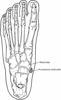
- Navicular bone and accessory navicular location in the foot.
- The accessory navicular (os navicularum or os tibial externum) is an additional(extra) bone or piece of cartilage placed at the internal aspect of the foot area simply above the arch. It is integrated with the back of the tibial tendon, which attaches to this area and can cause accessory navicular syndrome.
- An accent navicular is congenital (gift at birth). It isn’t always a part of ordinary bone shape and consequently isn’t always found in maximum human beings.
What Is Accessory Navicular Syndrome?
People who have an accessory navicular regularly are blind to the circumstance if it reasons no problems. whenever many human beings with this extra bone expand a painful circumstance referred to as accessory navicular syndrome while the bone and posterior aspect of a tibial tendon are aggravated. This could end result from any of the following
- Trauma, as in a foot area or ankle sprain
- Chronic inflammation from footwear or different shoes rubbing towards the more bone
- Excessive interest or overuse
Classification
The Geist type divides the accessory navicular bones into 3 types.
- Type 1: An os tibiale externum is a 2-three mm sesamoid bone inside the distal aspect of the posterior tibialis tendon. Usually asymptomatic.
- Type 2: Like a heart shape or triangular shape formed ossicle measuring almost twelve Milli meters, which illustrates a secondary ossification middle linked to the navicular bone tuberosity with the aid of using a one–two mm layer of fibrocartilage or hyaline cartilage. Portions of the posterior aspect of the tibialis tendon from time to time insert onto the accent ossicle, which could motivate dysfunction, and therefore, symptoms
- Type 3: A cornuate type of navicular bone depicts a large(big) measurement of the navicular bone tuberosity, which could else secondary constitute a fused type two accent bone. Occasionally symptomatic because of bunion type of formation.
What Causes An Accessory Navicular Syndrome?
- The majority of human beings with accent naviculars do now no longer have symptoms. However, this extraneous bone can worsen the posterior tibial tendon, causing aches and swelling. People who do experience accent navicular syndrome expand aches because of overuse, direct trauma, or chronic inflammation from shoes.
- While nobody is aware of what reasons accent navicular syndrome, it’s miles a congenital condition. Experts suppose that it can be a result of an incomplete fusion of the bone and connective tissue at some stage in development. It will also be a result of a strange separation of the bone and connective tissue.
What Are The Symptoms Of Accessory Navicular Syndrome?
- Accessory navicular symptoms often appear during adolescence. However, in some cases symptoms can start in adulthood. some patients present with a prominent bony area on the medial side, or inner side, of the foot or arch. They may also experience pain in the foot, swelling, and occasional redness. These symptoms are generally aggravated by walking time or standing for long periods of time.
- If you’ve got got an accessory navicular, you could now no longer be conscious which you have when you have no symptoms. Pain, swelling, throbbing, and redness with inside the arch of the foot are symptoms and symptoms of aggravation. You can also additionally observe a seen bony location at the mid-foot. Sources of accent navicular syndrome might also additionally consist of the following:
- difficulty in raising the foot
- medial arch pain
- swelling medial aspect of the mid foot
- Trauma, as in a foot or ankle muscles sprain
- Chronic infection from footwear or different shoes rubbing towards the more bone
- Excessive activity or overuse
How Is An Accessory Navicular Diagnosed?
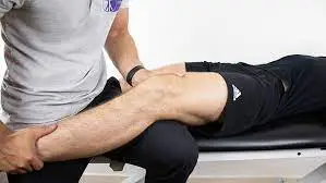
- The doctor asks about the patient’s signs and symptoms to diagnose accessory navicular syndrome and examines the foot area for pores and skin inflammation or swelling.The practitioner might also additionally press on the bony prominence to evaluate the place for discomfort. Foot structure, muscle strength, joint motion, and the manner the affected person walks can also be evaluated.
- X-rays are normally ordered to confirm the diagnosis. If there is ongoing ache or inflammation, an MRI or different advanced imaging assessments can be used to similarly examine the condition.
Nonsurgical Treatment Approaches
The determine of nonsurgical therapy approach for accessory navicular syndrome is to reduce the accessory navicular symptoms. The following can be used:
- Immobilization: Placing the ankle area in a castor or removable walking boot gives the affected area a sense of relaxed muscle area and reduces inflammation.
- Ice. To decrease swelling, a bag of ice protected with a skinny towel is implemented to the affected region. Do now no longer position ice immediately at the skin.
- Medications. Oral non-steroidal anti-inflammatory drugs (NSAIDs), together with ibuprofen, can be prescribed. In a few cases, oral or injected steroid medicinal drugs can be utilized in aggregate with immobilization to decrease pain and inflammation.
- Physical therapy. Physical therapy can be prescribed, which includes activities and treatment to strengthen the muscle mass and reduce inflammation. The exercise may also help prevent the recurrence of the symptoms.
- Orthotic devices. Custom orthotic devices that shape the shoe provide support for the arch and can play a function in preventing future symptoms.
Physiotherapy Treatment
Soleus Stretch Standing
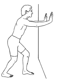
- Stand upright with the affected leg in the back of you
- Bend the again leg even as keeping your heel on the ground
- Stop and hold when you feel a pull at the again of your leg
- Hold for forty-five seconds
- Repeat 3-four times daily.
Foot Stretch
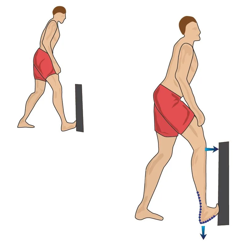
- Stand upright together along with your foot towards a step and toes extended
- Keep your heel on the ground and move your knee over your foot
- Stop and hold when you feel a pull at the again of your leg and the sole of your foot
- Hold for forty-five seconds
- Repeat 3-four times daily.
Posterior Tibial Muscle Strengthening (Band)
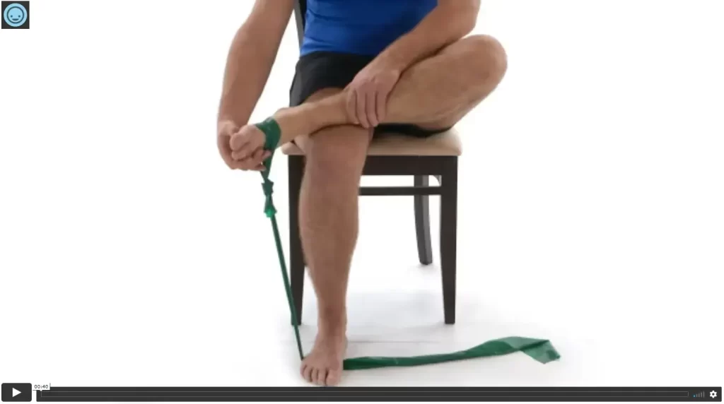
- In a seated position, place the affected ankle over the other knee
- Place a band around each foot
- Point the toes of the affected foot and raise them up in opposition to the resistance of the band
- Slowly go back to your beginning role to finish one repetition
- Repetitions: 15
- Sets: 3
- Frequency: Once daily
Posterior Tibial Muscle Strengthening (Ball)
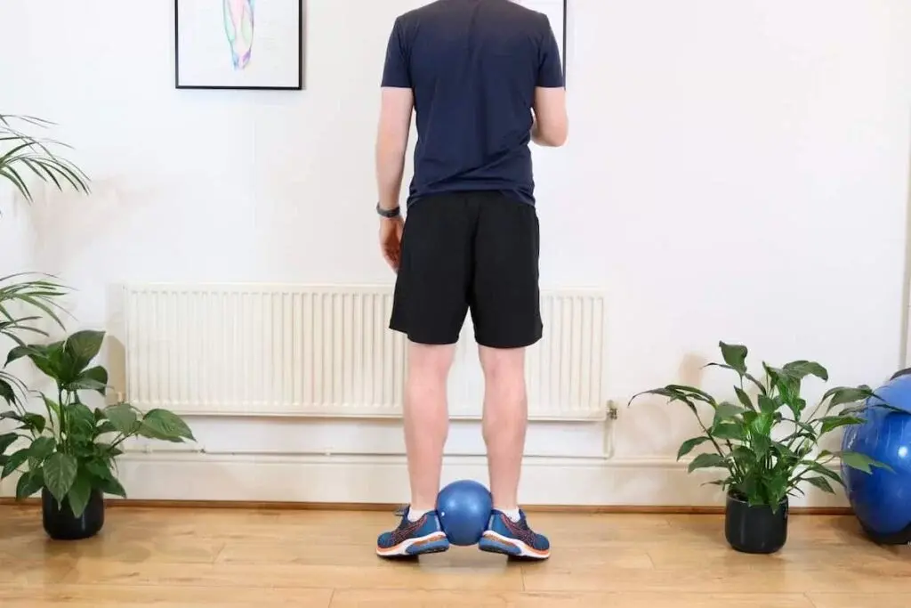
- Standing, place a ball among your heels and turn your feet outwards
- Raise your heels from the ground at the same time as squeezing the ball together along with your heels
- Once you attain the top, slowly go back to your beginning role to finish one repetition
- Repetitions: 15
- Sets: 3
- Frequency: Once daily
Single-Leg Balance: Eyes Open

- Stand on one leg together along with your foot flat on the ground
- Hold your contrary leg out in the front
- Focus on a factor in front of you to assist with stability
- Hold for 60 seconds
- Repeat 3-four times daily.
Single-Leg Balance: Wobble Cushion

- Stand on one leg together along with your foot on a wobble cushion on the floor
- Hold your opposite leg out in the front
- Focus on a factor in front of you to assist with stability
- Hold for 60 seconds
- Repeat 3-four times daily.
Surgical Treatment
- If accessory navicular syndrome cannot always be corrected by nonsurgical methods, additional navicular surgical procedures may be required to remove extraneous bone fragments. Because the accessory bone is not always necessary for foot function, its removal often relieves symptoms after recovery. In many cases, the accessory bone must be removed and also the posterior part of the tibial tendon must be reattached to the foot joint only. Additional navicular bone surgery relieves painful symptoms in 90% of cases.
Why choose a foot and ankle surgeon?
- Foot and ankle surgeons are the main professionals in foot and ankle care today. As medical doctors of podiatric medicine – additionally referred to as podiatrists, DPMs, or occasionally “foot and ankle medical doctors” – they’re the board-licensed surgical professionals of the podiatric doctors. Ankle joint and foot joint surgeons have more education and training specific to the foot joint and ankle joint than many other doctors.
- Foot joint and ankle joint surgeons deal with all situations affecting the foot joint and ankle joint from the easy to the complex in patients of every age inclusive of Accessory Navicular Syndrome. Their intensive education and specific training qualify foot joint and ankle joint doctors to perform a wide range of surgeries inclusive of any surgical operation that can be indicated for Accessory Navicular Syndrome.
FAQs
A visible bony spur on the inside of the foot
Redness and swelling of the bony spur
Pain in the foot area around the bony spur especially during or after some activity
It is when there is an excess segment of bone or cartilage in the foot region next to the navicular bone causing pain.
There are 3 types of accessory naviculars: Type 1 is an ossicle in the substance of the posterior tibial
tendon; Type 2 forms a synchondrosis with the navicular bone; and Type 3, “the cornuate navicular,”
represents the possible end stage of Type 2
This painful situation is referred to as accessory navicular syndrome. Accessory navicular syndrome (ANS) can cause significant aches in the mid-foot and arch, especially with activity. Redness and swelling may develop over this bony prominence, as well as extreme sensitivity to pressure.
This congenital defect (gift at birth) is thought to occur during improvement while the bone is
calcifying. Because this accessory part of the bone and the navicular never quite develop together, it is
believed that, over time, the excessive movement among the 2 bones outcomes in pain.