Sudeck’s Atrophy (Complex Regional Pain Syndrome)
Table of Contents
What is Sudeck’s atrophy?
Complex regional pain syndrome (CRPS), also known as reflex sympathetic dystrophy (RSD), is a disorder of a portion of the body, usually, the arms or legs, which manifests as pain, swelling, limited range of motion, and changes to the skin and bones. It may initially affect one limb and then spread throughout the body; 35% of affected people report symptoms throughout their whole bodies. There are multiple names for this disease, as well as two subtypes. Type I (also called reflex sympathetic dystrophy) refers to CRPS without evidence of a specific peripheral nerve injury. Type II refers to when there is specific evidence of a nerve injury.
Signs & Symptoms of Sudeck’s Atrophy
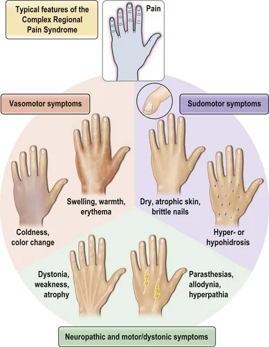
The symptoms are variable and will present themselves differently from patient to patient. The main symptoms are –
- a generalized burning pain
- changes in the skin, which may become shiny
- swelling
- more perspiration than usual
- muscle wastage
- stiffness in the affected part
- increased sweating
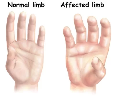
Stages of Complex Regional Pain Syndrome (CRPS)
Previously it was considered that CRPS had three stages; it is now thought, instead, that such individuals are likely to have one of the three following types of disease progression:
- “Stage” one is characterized by severe, burning pain at the site of the injury, muscle spasms, joint stiffness, restricted mobility, rapid hair and nail growth, and vasospasm. The vasospasm is that which causes the changes in the color and temperature of the skin. Some may experience hyperhydrosis (increased sweating). In mild cases, this stage lasts a few weeks, in which it can subside spontaneously or respond rapidly to treatment (physical therapy, pain specialist).
- “Stage” two is characterized by more intense pain. Swelling spreads, hair growth diminishes, nails become cracked, brittle, grooved, and spotty, osteoporosis becomes severe and diffuse, joints thicken, and muscles atrophy.
- “Stage” three is characterized by irreversible changes in the skin and bones, while the pain becomes unyielding and may involve the entire limb. There is marked muscle atrophy, severely limited mobility of the affected area, and flexor tendon contractions (contractions of the muscles and tendons that flex the joints). Occasionally the limb is displaced from its normal position, and marked bone softening and thinning is more dispersed.
Causes of Sudeck’s Atrophy
Investigators estimate that 2–5% of those with peripheral nerve injury, and 13-70 percent of those with hemiplegia, will develop CRPS. In addition, some studies have indicated that cigarette smoking was strikingly present in patients and is statistically linked to RSD. This may be involved in its pathology by enhancing sympathetic activity, vasoconstriction, or some other unknown neurotransmitter-related mechanism. This hypothesis was based on a retrospective analysis of 53 patients with RSD, which showed that 68% of patients and only 37% of controls were smokers. The results are preliminary and are limited by their retrospective nature.7% of people who have CRPS in one limb later develop it in another limb.
PATHOPHYSIOLOGY
Complex regional pain syndrome is a multifactorial disorder with clinical features of neurogenic inflammation (swelling in the central nervous system), nociceptive sensitization (which causes extreme sensitivity or allodynia), vasomotor dysfunction (blood flow problems which cause swelling and discoloration), and maladaptive neuroplasticity; CRPS is the result of an “inappropriate response to tissue injury”.
The “underlying neuronal matrix” of CRPS is seen to involve cognitive and motor as well as nociceptive processing; pinprick stimulation of a CRPS affected limb was painful (mechanical hyperalgesia) and showed a “significantly increased activation” of not just the S1 cortex (contralateral), S2 (bilateral) areas, and insula (bilateral) but also the associative-somatosensory cortices (contralateral), frontal cortices, and parts of the anterior cingulate cortex. In contrast to previous thoughts reflected in the name RSD, it appears that there is reduced Sympathetic Nervous System outflow, at least in the affected region (although there may be sympatho-afferent coupling).
Wind-up (the increased sensation of pain with time) and central nervous system (CNS) sensitization are key neurologic processes that appear to be involved in the induction and maintenance of CRPS.
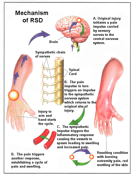
The pathophysiology of complex regional pain syndrome has not yet been defined; there is conjecture that CRPS, with its variable manifestations, could be the result of multiple pathophysiologies.
DIAGNOSIS
There is no biological test to highlight Sudeck’s disease. The physician makes the diagnosis based primarily on physical findings and history.
- Temperature measurements: temperature measurement generated by the body surface
- Measuring the secretion of sweat
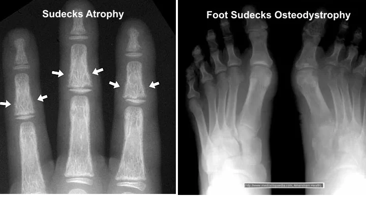
- Radiography, CT, MRI: to highlight a bone decalcification.
- Blockages friendly: using anesthesia can reduce sympathetic activity, this method allows obtaining diagnostic information and therapeutic action, insofar as Sudeck’s disease depends on the sympathetic system.
- Scintigraphy in three phases: review specific nuclear medicine
- The pain and swelling in the limbs may also come from other illnesses, so it is important to eliminate them. (venous thrombosis, lymphedema as an accumulation of fluid and lymphatic system disorders, lymph node involvement in cases of breast cancer).
The International Association for the Study of Pain (IASP) lists the diagnostic criteria for complex regional pain syndrome I (RSDS) as follows:
- The presence of an initiating noxious event or a cause of immobilization
- Continuing pain, allodynia (perception of pain from a nonpainful stimulus), or hyperalgesia (an exaggerated sense of pain) disproportionate to the inciting event
- Evidence at some time of edema, changes in skin blood flow, or abnormal sudomotor activity in the area of pain
- The diagnosis is excluded by the existence of any condition that would otherwise account for the degree of pain and dysfunction.
According to the IASP, CRPS II (causalgia) is diagnosed as follows:
- The presence of continuing pain, allodynia, or hyperalgesia after a nerve injury, not necessarily limited to the distribution of the injured nerve
- Evidence at some time of edema, changes in skin blood flow, or abnormal sudomotor activity in the region of pain
- The diagnosis is excluded by the existence of any condition that would otherwise account for the degree of pain and dysfunction.
Treatment of Sudeck’s Atrophy
The goal of treatment is the preservation of the normal functionality of the affected body part, that is to say, the mobility of the extremities. Treatment requires patience from the patient and physician. It is very difficult to influence the course of the disease and the psychological burden caused by the pain is very important.
Medical treatment:
Tentative evidence supports the use of bisphosphonates, calcitonin, and ketamine. Doing nerve blocks with guanethidine appears to be harmful. Evidence for sympathetic nerve blocks generally is insufficient to support their use. Intramuscular botulinum injections may benefit people with symptoms localized to one extremity.
- Ketamine
Ketamine, a dissociative anesthetic, appears promising as a treatment for complex regional pain syndrome. It may be used in low doses if other treatments have not worked. No benefit on either function or depression, however, has been seen. - Bisphosphonate treatment
Very limited data reviewed showed that bisphosphonates have the potential to reduce pain associated with bone loss in patients with CRPS I.
Surgery
It is required when medication and conservative treatment are failed and is mostly used in severe CRPS. The following are the procedures:
- Spinal cord stimulators
- Sympathectomy
- Amputation
Physiotherapy Treatment
It is important that the patient actively participates and takes responsibility during treatment.
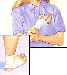
- First of all, to rest; possibly splinting
- Elevation of the affected
- Cool slightly (NOT ice), carbon dioxide baths costs
- Manual lymphatic drainage
- Electrotherapy, IFT, TENS (transcutaneous electrical nerve stimulation: inhibition of conduction of pain due to nerve stimulation by electrical current)
- Massages of connective tissue
- Mobilization in the chronic phase
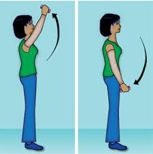
- Carefully active movement of the affected limb
- Occupational therapy to maintain mobility
- Specific training with specialists
- Learning relaxation techniques
- Aquatic therapy allows activities to be performed with decreased weight bearing on the lower extremities
- Mirror therapy
- Gradual weight bearing
- Regular Gradual Stretching exercise in Pain-free mode
Other treatments:
- Whirlpool bath/ contrast baths
- Vocational and recreational rehabilitation
- Psychological therapies: cognitive-behavioral therapy (CBT), operant conditioning (OC), counseling, pain education, and relaxation techniques
- Acupuncture & electroacupuncture
- Tactile sensory discrimination training
- Ultrasound therapy
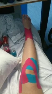
- Kinesio taping
FAQs
Reflex sympathetic dystrophy, Sudeck’s atrophy, shoulder-hand syndrome, or causalgia were earlier names for this illness.
The area is initially painful, throbbing, red, heated, and swollen. After a few weeks or months, the area may turn cold, mottled, glossy, stiff, and frequently have underlying osteoporosis. Later, when the muscles continue to atrophy and possibly contract, the pain persists.
The “acute bone atrophy” that is frequently a part of the “pathological syndrome of the hand” is caused by circulatory issues that develop on traumatized hands or hands that have been injured “at a distance,” especially when nerve damage is present.
Although it can happen at any age, it is more common in women between the ages of 40 and 60, despite the fact that young people are being diagnosed with it more frequently. Strong analgesic and neuromodulatory painkillers are used to regulate it; some of the latter are also used to treat psychiatric problems, epilepsy, etc.

One Comment