Sciatica: Physiotherapy Treatment
Table of Contents
What is Sciatica Pain?
Sciatica is a condition that can lead to pain in the low back and legs. It occurs when pain travels along the path of the sciatic nerve. This nerve starts in your lower spine, and travels through your hip and buttock and down the back of your leg to your foot.
Sciatica is not so much a condition but actually a symptom. Mostly sciatica affects one side of Body, but it can also affects bilaterally simultaneously.
Sciatica most commonly occurs when a tight piriformis muscle, disk bulge, arthritis of low back on the spine or narrowing of the spine (spinal canal stenosis) compresses part of the nerve. This causes radiating pain and often tingling numbness in the affected leg.
Sciatica pain can be cured with non-operative treatments in a few weeks. If you have severe sciatica pain that’s associated with significant leg weakness or bowel or bladder changes might be suggested for surgery.
ANATOMY OF SCIATIC NERVE:
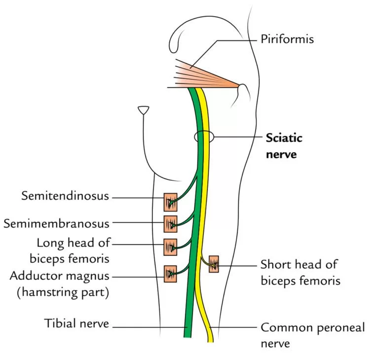
The sciatic nerve is formed from the L4 to S3 segments of the sacral plexus, a collection of nerve fibres that emerge from the sacral part of the spinal cord. The fibres unite to form a single nerve in front of the piriformis muscle. The nerve passes beneath piriformis and through the greater sciatic foramen, exiting the pelvis. it travels down the posterior thigh to the popliteal fossa.
The nerve travels in the posterior compartment of the thigh behind (superficial to) the adductor magnus muscle, and is itself in front of (deep to) one head of the biceps femoris muscle. At the popliteal fossa, the nerve divides into its two branches. The tibial nerve, which travels down the posterior compartment of the leg into the foot. The common peroneal nerve (also called the common fibular nerve), which travels down the anterior and lateral compartments of the leg into the foot
Pathophysiology:
Sciatica is generally caused by the compression of lumbar nerves L4 or L5 or sacral nerve S1. Less commonly, sacral nerves S2 or S3 or compression of the sciatic nerve itself may cause sciatica. In 90% of sciatica cases, this can occur as a result of a spinal disc bulge or herniation. Intervertebral spinal discs consist of an outer anulus fibrosus and an inner nucleus pulposus. The anulus fibrosus forms a rigid ring around the nucleus pulposus early in human development, and the gelatinous contents of the nucleus pulposus are thus contained within the disc. Discs separate the spinal vertebrae, thereby increasing spinal stability and allowing nerve roots to properly exit through the spaces between the vertebrae from the spinal cord. As an individual age, the anulus fibrosus inevitably weakens and becomes less rigid; thus, it is subsequently more likely to tear.
When there is a tear in the anulus fibrosus, the nucleus pulposus may extrude through the tear and press against spinal nerves within the spinal cord, cauda equina, or exiting nerve roots, causing inflammation, numbness, or excruciating pain. Inflammation of spinal tissue can then spread to adjacent facet joints and cause facet syndrome, which is characterized by lower back pain and referred pain in the posterior thigh.
Other causes of sciatica secondary to spinal nerve entrapment include the roughening, enlarging, or misalignment (spondylolisthesis) of vertebrae, or disc degeneration that reduces the diameter of the lateral foramen through which nerve roots exit the spine. When sciatica is caused by compression of a dorsal nerve root, it is considered lumbar radiculopathy or radiculitis when accompanied by an inflammatory response. Sciatica-like pain prominently focused in the buttocks can also be caused by compression of peripheral sections of the sciatic nerve usually from soft tissue tension in the piriformis or related muscles.
Sciatica Symptoms:
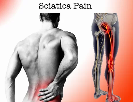
- hot pain in the buttock
- aching in the buttock, back of the thigh (hamstring) and calf
- pain in the ankle and foot
- pins and needles
- increased pain when lifting, straining, or coughing
- loss of power to the muscles of the leg and foot.
Sciatica Causes:
Sciatica is caused by irritation of the sciatic nerve. Usually, there is no specific injury that is related to the onset of sciatica. Occasionally, the pain will suddenly begin after lifting something heavy or moving quickly. The following are the causes of sciatica:
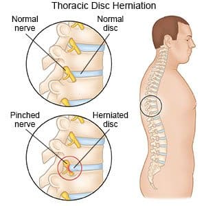
A herniated disc (sometimes called a slipped disc): Disc herniation is the most common cause of sciatica. When a disc herniates near the spinal nerve roots that form the sciatic nerve, it can cause pressure on the nerve, or irritation, which results in the symptoms of sciatica. Discs are the cushions between the bones in the back. They act like “shock absorbers” when we move, bend, and lift.
They are the size and shape of checkers. There is a tough ring around the outside of each disc and a thick jellylike center inside (called a nucleus pulposus). If the outer edge of the disc ruptures, the center can push through and put pressure on the sciatic nerve, leading to the pain of sciatica (referred to as a herniated nucleus pulposus).
Lumbar spinal stenosis, a narrowing of the canal that contains the spinal cord: With age, the bone can overgrow and put pressure on the sciatic nerve. Many people with spinal stenosis have sciatica on both sides of the back.
Lumbar Spondylolisthesis is a condition in which one backbone has slipped forward or backward over another backbone, which can result in pressure on the sciatic nerve. A pinched or stretched sciatic nerve
Piriformis syndrome can cause the sciatic nerve to become trapped deep in the buttock by the piriformis muscle. The symptoms of piriformis syndrome are the same as those of sciatica.
Sciatica can also be caused by the nerves being pinched by osteoarthritis and fractures due to osteoporosis.
Sciatica can also be caused by other effects of aging, such as osteoarthritis and fractures due to osteoporosis.
Many women experience sciatica during pregnancy.
Diagnosis:
X-ray: An X-ray of your spine may reveal an overgrowth of bone (bone spur) that may be pressing on a nerve.
MRI: This procedure uses a powerful magnet and radio waves to produce cross-sectional images of your back. An MRI produces detailed images of bone and soft tissues such as herniated disks. During the test, you lie on a table that moves into the MRI machine.
CT scan: When a CT is used to image the spine, you may have a contrast dye injected into your spinal canal before the X-rays are taken — a procedure called a CT myelogram. The dye then circulates around your spinal cord and spinal nerves, which appear white on the scan.
Electromyography (EMG): This test measures the electrical impulses produced by the nerves and the responses of your muscles. This test can confirm nerve compression caused by herniated disks or narrowing of your spinal canal (spinal stenosis).
PHYSICAL EXAMINATION :
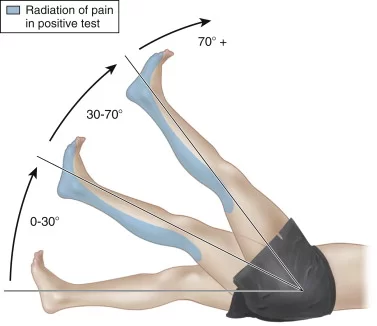
Straight leg raise test (SLR Test ) :
the patient is positioned in supine without a pillow under his/her head, the hip medially rotated and adducted, and the knee extended. The clinician lifts the patient’s leg by the posterior ankle while keeping the knee in a fully extended position. The clinician continues to lift the patient’s leg by flexing at the hip until the patient complains of pain or tightness in the back or back of the leg.A positive test is considered when the patient reports reproduction of pain at 40 degrees of hip flexion or less.
Slump test :
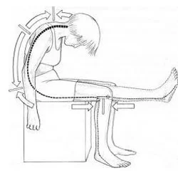
To begin the test, have the patient seated with hands behind the back to achieve a neutral spine. The first step is to have the patient slump forward at the thoracic and lumbar spine. If this position does not cause pain, have the patient flex the neck by placing the chin on the chest and then extending one knee as much as possible.
If extending the knee causes pain, have the patient extend the neck into neutral. If the patient is still unable to extend the knee due to pain, the test is considered positive.
If extending the knee does not cause pain, ask the patient to actively dorsiflex the ankle. If dorsiflexion causes pain, have the patient slightly flex the knee while still dorsiflexing. If the pain is reproduced, the test is considered positive.
Repeat the test on the opposite side.
Femoral nerve tension test:
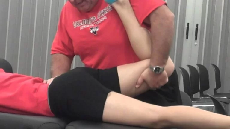
The patient is prone symmetrically on the bed, the clinician places one hand on the patient’s pelvis to prevent movement and feel for any compensations, while the other hand flexes the involved knee as much as possible and maintains the position for 45 seconds. The test is positive if the patient experiences anterior thigh pain. This test is usually positive for L2-L3 and L3-L4 (high lumbar) protrusions, slightly positive or negative in L4–L5, and negative in cases of lumbosacral protrusion.
Sciatica Treatment
Medical Treatment:
Over-the-counter pain relievers can provide short-term relief from sciatica. Acetaminophen and nonsteroidal anti-inflammatory drugs (NSAIDs), such as aspirin, ibuprofen, and naproxen are options. Your doctor may give you a steroid injection to further reduce the inflammation.
Steroid injections:
In some cases, your doctor might recommend injection of a corticosteroid medication into the area around the involved nerve root. Corticosteroids help reduce pain by suppressing inflammation around the irritated nerve. The effects usually wear off in a few months. The number of steroid injections you can receive is limited because the risk of serious side effects increases when the injections occur too frequently.
Surgery
Laminectomy:
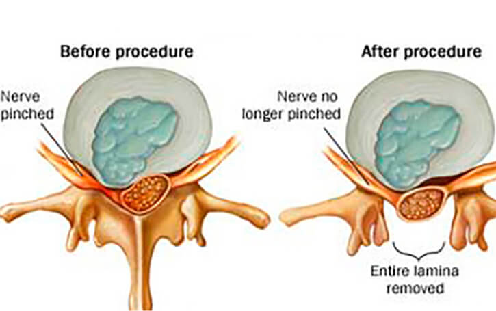
The bone that curves around and covers the spinal cord (lamina), and the tissue that is causing pressure on the sciatic nerve are removed.
PHYSIOTHERAPY TREATMENT :
Physiotherapy treatment mainly depends upon the diagnosis of sciatica and also symptomatic pain-relieving electrotherapy modalities.
IFT (Interferential Therapy) :
IFT are used to relieve sciatica-referred pain where two electrodes are placed at the start of the nerve roots and the other two electrodes are used at the leg, IFT is highly used by physiotherapists in their clinic for sciatica.
Lumbar Traction:
This Intermittent lumbar traction helps to relieve pressure on sciatic nerve roots and is widely used by Physiotherapists to relieve sciatica.
TENS (Transcutaneous Electrical Nerve Stimulation) :
TENS is also used to relieve sciatica referred to as pain. The use of two electrode pads placed on the skin starts on the sciatic nerve roots and TENS is a battery-operated device delivering a small current to them to produce a tingling sensation.
Ice and Heat:
There are steps you can take at home to ease the pain of sciatica. A heating pad or ice pack may be especially helpful. Apply the heat or ice for about 20 minutes every two hours.
Acupuncture:
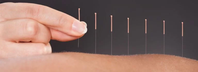
In acupuncture, the practitioner inserts hair-thin needles into your skin at specific points on your body. Some studies have suggested that acupuncture can help back pain, while others have found no benefit. If you decide to try acupuncture, choose a licensed practitioner to ensure that he or she has had extensive training.
Physiotherapy Exercise:
Exercises for sciatica
The following Exercise helps To Release Sciatic Nerve :
Knee to chest stretch :
Lie on your back on a mat or the carpet. Place a small, flat cushion or book under your head. Bend your knees and keep your feet straight and hip-width apart. Keep your upper body relaxed and your chin gently tucked in. Bend one knee up towards your chest and hold it with both hands. Hold for 20 to 30 seconds with controlled deep breaths.
Sciatic Mobilizing Stretch:
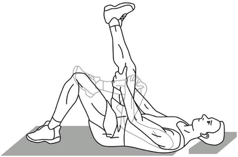
Sciatic Mobilizing Stretch
Lie on your back. Place a small, flat cushion or book under your head. Bend your knees and keep your feet straight and hip-width apart. Keep your upper body relaxed and your chin gently tucked in. Bend one knee up towards your chest. Hold the back of your upper leg with both hands, then slowly straighten the knee. Hold for 20 to 30 seconds, taking deep breaths. Bend the knee and return to the starting position.
Supine Piriformis Stretch:
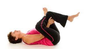
Lie on the floor with the affected leg crossed over the other leg at the knees and both legs bent. Slowly pull your lower knee towards your shoulder on the same side of your body until you feel the stretch. Hold for 30 seconds, then slowly go back to the starting position.
Transversus Abdominus:
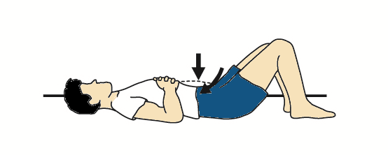
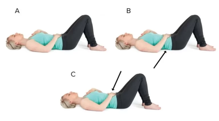
The patient pulls his belly in and up at the navel without moving the rib cage, pelvis, or spine. Contraction intensity: 30 to 40% of the maximum voluntary contraction
Back extensions:
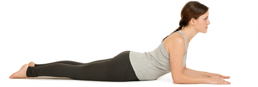
Lie on your front and rest on your forearms with your elbows bent at your sides. Look towards the floor and keep your neck straight.
Keeping your neck straight, arch your back up by pushing down on your hands. You should feel a gentle stretch in the stomach muscles. Breathe and hold for 5 to 10 seconds. Return to the starting position.
Sitting spinal stretch :
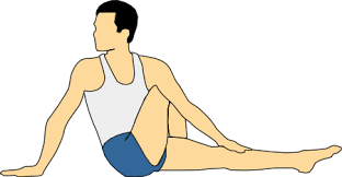
Sit on the ground with your legs extended straight out with your feet flexed upward.
Bend your right knee and place your foot flat on the floor on the outside of your opposite knee.
Place your left elbow on the outside of your right knee to help you gently turn your body toward the right.
Hold for 30 seconds and repeat three times, then switch sides.
Pelvic Tilt :
Lie on your back with your knees bent and your feet flat on the floor. Inhale. Flatten your back against the floor by tightening your abdominal muscles and bending your hips slightly.
Exhale. As you exhale, move your hips upward without lifting your rear end to remove the natural curve. As you inhale, slowly return to the starting position.
Lower Trunk Rotations :
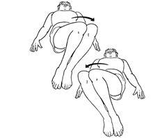
Lie on your back with both knees bent upright and both feet flat on the floor (called the hook lying position).
While holding both knees together, rotate your knees to one side and hold for 3 to 5 seconds. You will feel a gentle stretching sensation in the opposite side of your lower back and hip area.
Next, contract your abdominal muscles and rotate both knees to the opposite side and hold for 3 to 5 seconds.
Repeat up to 10 times on each side.
Hamstring stretching Exercise :
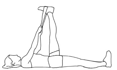
Lie on the back, supporting the thigh with the hand or with a towel wrapped around it. Slowly straighten the knee until a stretch is felt in the back of the thigh, trying to get the bottom of the foot to face the ceiling, one leg at a time. Hold the position initially for 10 seconds, and gradually work up to 20 to 30 seconds.
DO’S and DON’T:
- sit correctly when using a computer
- lose weight if you’re overweight
- Carry on with your normal activities as much as possible
- regular back stretches (Improve Back muscle flexibility )
- start gentle exercise as soon as you can – anything that gets you moving can help (Strengthen Back muscle)
- hold heat packs to the painful areas – you can buy these from pharmacies
- ask your pharmacist about painkillers that can help – paracetamol on its own is unlikely to relieve your pain
- Exercise regularly.
- Maintain good posture.
- Bend at the knees to lift heavy objects.
- do not sit or lie down for long periods – even if moving hurts, it’s not harmful and can help you get better faster
- do not use hot water bottles to ease the pain – you could scald yourself if your skin is numb.



13 Comments