Leg Length Test
The leg length test is a simple yet informative diagnostic examination frequently used in the fields physical therapy, and orthopedics. It serves as an essential tool for assessing the structural alignment and biomechanics of the lower limbs, particularly the legs, and can provide valuable insights into a patient’s musculoskeletal health.
Table of Contents
Definition/Description
- Inequality in leg length is frequently related to compensatory gait abnormalities and can lead to lower extremities and lumbar spine degenerative arthritis. Leg-length discrepancy (LLD) patients may also have angular and torsional deformities, as well as soft tissue contractures of the ipsilateral or contralateral limbs, all of which can impede functional leg lengths.
- Flexion contractures around the knee and hip, for example, may cause the leg to look shorter, but abduction contractures of the hip and equinus deformity of the ankle tend to effectively extend the afflicted extremity. Aside from clinical examination, many imaging methods have been reported to measure LLD. To adequately treat a patient with uneven leg lengths or associated symptoms, it is critical to apply suitable clinical procedures and imaging modalities for assessing the LLD.
- Leg length discrepancy (LLD), also known as anisomelia, is a disorder that causes the lengths of the paired lower extremity limbs to be significantly variable. For many years, scholars and physicians have been discussing the topic of leg length discrepancy (LLD). The presence of LLD has been recognized but there is little agreement on its many aspects, such as the extent of LLD considered clinically significant, the prevalence, reliability, The reliability and validity of the measurement methods, the influence of LLD on function, and its involvement in diverse neuromusculoskeletal disorders are all investigated.
Leg Length Disparity Types
Leg length disparity (LLD) may be classified into two categories.
Anatomical
Inequality in structural limb length. Congenital problems are mild developmental defects that occur at birth or during childhood, whereas acquired conditions include trauma, fractures, orthopedic degenerative illnesses, and surgical disorders such as joint replacement. A radiographic-based study of the prevalence of LLD indicated that 90% of the normal population had some variation in bony leg length, with 20% having a variance of more than 9 mm.
Functional
- Shortening that has no structural basis. It is a unilateral lower extremity asymmetry with no shortening of the lower limb’s osseous components. A change in lower limb mechanics, such as joint contracture, static or dynamic mechanical axis misalignment, muscle weakening, or shortening, causes FLLD.
A non-functional examination, such as radiography, cannot identify these poor mechanics. FLLD can occur as a result of aberrant hip, knee, ankle, or foot mobility in any of the three planes of motion. - It is clear that research involving persons with long-standing true LLD may deal with more LLD than research involving those who have been subjected to artificial or induced LLD. This is logical since given enough time, most people could lower the energy and mechanical costs of LLD. Furthermore, younger people are better able to adjust to bigger LLD than older people (which makes sense given that gait patterns change significantly between old and young people and older people have more trouble learning novel motor tasks). The individual’s degree of exercise appears to have a factor as well. Individuals who spend most of their day on their feet or who participate in sports who are more active appear to be more vulnerable to LLD than those who are less active.
Etiological Factors
- True LLD
- Idiopathic developmental disorders
- Fracture
- Epiphyseal endplate trauma before skeletal maturity
- Degenerative conditions
- Legg-Calvé-Perthes Syndrome
- Neoplastic or cancerous alterations
- Infections
- LLD is functional soft tissue shortening
- Joint contractures
- Laxity of the ligaments
- Misalignments of the axes
- Foot biomechanics (excessive ankle pronation, for example)
Purpose
- The goal of this test is to determine the leg length discrepancy (LLD).
- Leg length differences are often divided into two categories: genuine and functional.
- True LLD are those with a genuine bony asymmetry midway between the femoral head and the ankle mortise.
- Functional LLD is those that occur as a physiological reaction to altered mechanics across the kinetic chain from the foot to the lumbar spine, giving the sense of a short leg despite the absence of actual asymmetry in bone length.
Technique
Direct Method:
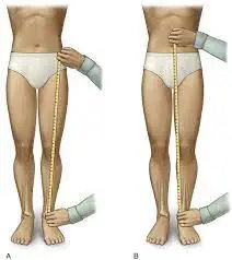
- In the stand, limb length is measured with a tape measure between two specified locations. The anterior iliac spine and the medial malleolus are two common sites, as are the anterior inferior iliac spine and the lateral malleolus.
- However, be aware because there has been a lot of criticism and controversy about the accuracy of tape measure methods. Keep the following subjects and potential faults in mind if you use this method:
- Always use the mean of at least two or three measurements.
- Compare measurements between two or more clinicians if possible.
- Iliac asymmetries may cover up or amplify a limb length disparity.
- Unilateral variations in the long axis of the lower limb (e.g., Genu varum,…) may conceal or exaggerate a limb length disparity.
- The asymmetrical location of the umbilicus
- Joint contractures
Indirect Method
- In standing, palpation of bony landmarks, most often the iliac crests or anterior iliac spines. These approaches involve assessing whether or not bone landmarks are at (the horizontal) level and whether or not there is a limb length inequality.
The best (most accurate and exact) clinical approach to detect limb inequality appears to be palpation and visual evaluation of the iliac crest (or ASIS) together with the use of to alter the level of the iliac crests (or ASIS), place blocks or book pages of specified thickness beneath the shorter leg. - It Considers that asymmetric pelvic rotations in planes other than the frontal plane may indicate limb length discrepancy. A study of the literature recommends that while using the block correction approach, the greater trochanter major and as many pelvic landmarks as possible should be palpated and compared (left trochanter with right trochanter).
The PALM (Palpation Meter)
- The PALM is reliable and accurate equipment for determining pelvic height discrepancy. It is a simple and cost-effective alternative to radiographic measuring.
- Measurement Procedure: Two tape strips were laid 15cm apart on the ground. The tape strips indicate where patients have to put their feet on the floor. Patients are instructed to take 10 steps while aligning the medial borders of their feet with the outside of the tape strips. Patients must stand fully upright (no bending of the knees, hips, or spine). The PALM is located on the iliac crest’s most superior portion. The distance between the caliper heads is measured to the closest millimeter, while the angle of inclination is measured to the nearest half-degree. The inclinometer ball has been designed to travel toward the shorter limb’s side.
Standing on Blocks
- The patient is standing with her feet 10 cm apart, her knees extended, and her weight evenly distributed on both feet.
The doctor places his or her hands on one of the bilateral anatomical structures listed below: Spina iliaca posterior superior, Spina iliaca anterior superior, or crista iliaca left and right. If there is a length inequality, the physician visually examines it and inserts a 0.5% wooden board beneath the foot of the shorter side. - Continue to place thicker planks beneath the shorter side until an equal length is achieved, with the thickness of the plank equal to the leg length difference.
- Although the accuracy of the clinician’s measures is critical for dependability, this technique has produced good outcomes in inter-examination findings between highly educated physicians and medical students. According to the research, confounding variables include pelvic asymmetry, poor foot placement, obesity, joint contractures, scoliosis, and faulty measurement.
Imaging Method :
A number of imaging techniques can be utilized, including:
- Plain Radiography
- Orthoroentogenogram
- Scanogram
- Teleoroentgenogram: a full-length standing AP radiograph in the lower extremity.
- Computed Radiography.
- Micro-dose Digital Radiography is a type of computer-aided imaging that significantly minimizes radiation exposure to patients when compared with conventional radiographic procedures.
- Ultrasound: This treatment employs an ultrasonic transducer to locate bone landmarks at the hip, knee, and ankle joints.
- An Anteroposterior (AP) scout view of the bilateral femurs and tibias is produced using a CT scanogram.
- MRI Scan: Images from the MRI were acquired using a T1 weighted spin echo sequence, and the best coronal images were chosen for standardized femur length measurement using the typical bone landmarks of the femoral head and medial femoral condyle.
The Role of LLD in Running
- Running biomechanics differ from walking biomechanics, as do the effects of LLD. Running has increased vertical oscillation and no double support, therefore weight is not divided between legs. Running has a 30% stance phase, whereas walking has a 60% stance phase.
- This causes three times the stress on the lower extremities as walking. The evidence on the impact of running is inconsistent, however, it is suggested that the effect increases thrice.
Associated Musculoskeletal Disorders
- Low Back Pain The existing body of literature is fairly unknown. LLD appears to have an effect on the lumbar spine, at least partially, by causing lumbar scoliosis.LLD has been shown to produce frontal pelvic obliquity, which results in scoliosis.
- Asymmetry of joint angles is suggested to predispose individuals to lumbosacral osteoarthritis.
- Further study will determine if LLD is only related to or an objective causative component in low back pain (LBP). The available literature is contradictory and inconclusive.
- Hip Pain A longer leg may be a risk factor for Osteoarthritis (OA). The femoral head contact/weight-bearing area decreases as length increases. (+10mm- 5%/ +50mm – 25,1%).
- When combined with greater tone in hip abductors induced by the prolonged distance between origin and insertion, the longer leg is put in danger.
- Fractures caused by stress The higher frequency of stress fractures in the longer leg’s tibia, metatarsals, and femur appears to be in line with the stronger forces transmitted through the longer leg indicated under hip discomfort.
- Others Trochanteric bursitis, patellar capacity, patellar joint incongruences, and peroneus longus myofascial pain syndrome appear to be associated.
Leg Length Discrepancy After Total Hip Arthroplasty
Clinical Signs
- Patients are classified into three categories based on how their symptoms include:
- Most frequently, patients complain of pain as a result of an imbalance in muscular forces at the hip, knee, and spine.
Quadriceps and hamstring muscle pain and tiredness in the longer leg (flexed knee syndrome). Patients bend their knees during stride to keep their pelvis level. - Instability or dislocation caused by component orientation; discomfort is frequently caused by hip and periarticular muscle exhaustion from avoiding subluxations.
- Correctable cause following Total Hip Arthroplasty (THA) – first postoperative obvious LLD. Periarticular muscle spasms, lumbosacral scoliosis, and pelvic obliquity are the most common early postoperative LLD symptoms.
- They are both capable of producing this early postoperative LLD. The physical effect is a tilted pelvis relative to the floor, with the surgical hemipelvis being pushed inferiorly and the contralateral hemipelvis being pulled superiorly, resulting in an apparent LLD despite equal lengths from the anterior superior iliac spines to the medial malleoli.
- Patient education, reassurance, physical therapy muscle methods, and potentially a temporary shoe lift are the mainstays of treatment for this condition. Most instances resolve after six months of surgery, so the patient should be comforted.
Physical Therapy Management
Non-surgical interventions are mostly utilized to correct functional leg length abnormalities. It is also used to describe the moderate form of limb length discrepancy.
Non-surgical Treatment
- Stretches the muscles of the lower extremities. Individually, the Tensor Fascia Latae, adductors, hamstring muscles, piriformis, and Iliopsoas are stretched, as are any other muscles in the kinetic chain that require stretching or strengthening.
- This non-surgical method is also associated with the usage of shoe lifts. These shoe lifts are made up of either a shoe insert (up to 10-20mm of correction) or a shoe sole buildup on the shorter leg (up to 30-60mm of correction). This lift therapy should be introduced gradually and in tiny steps. See the section below for further information.
- Several studies have looked into the use of shoe lifts to treat low back pain in individuals with LLD. Gofton achieved good results: the patients received significant or total pain alleviation that lasted from 3 to 11 years after the procedure. Helliwell also observed individuals in whom 44% had total pain reduction and 45% had moderate or significant pain relief. Friberg observed that after an average of 18 months, 157 (of 211) people with LBP who were treated with shoe lifts were symptom-free.
- However, the research on the use of lift therapy is unclear.
FAQs
The “true” leg length may be measured from the anterior superior iliac spine (ASIS) to the medial malleolus using a “direct” measurement using a tape measure. The “apparent” leg length is calculated by measuring the distance between the umbilicus and the medial malleolus. Morrissy RT, Weinstein SL, eds., reprinted with permission.
Leg lengths should be measured to see whether there is a discrepancy from one side to the other. It is critical to distinguish between genuine and perceived leg length deficiencies. A pelvic obliquity, contractures, or scoliosis may cause an apparent or functional leg length disparity.
Congenital limb differences cause some children’s legs or arms to develop at different rates. Major fractures, fractures that may not heal properly, bone infections such as osteomyelitis, and bone tumors or cysts are all possible causes of limb-length inequalities.
Leg length disparities are quite prevalent. In truth, many of us have a slight variation in our leg lengths. Most people, however, do not perceive the change and do not require therapy. Most doctors advocate having the disparity assessed if it is greater than 1.5 to 2.0 cm (5/8 of an inch).
Locate the bone junction where your hip connects to your leg. The anterior superior iliac spine, or ASIS, is where this occurs. Measure from this place to the bony joint on your ankle. Repeat on both sides and keep track of your observations.
Lower leg length (LLL), which is the distance between the length of a person’s foot and the knee, is one way for measuring height. This information may be used to compute total body length using a specific calculation.
Description. More than half of the population has a little discrepancy in leg length. These little changes seldom cause difficulties. However, for the tiny fraction of persons who have a leg length difference of more than 2 cm, the length discrepancy can have an impact on their well-being and quality of life.
If your leg length is unequal, you almost certainly have a spine misalignment. Even if your leg length difference is little, it causes an imbalance across your whole skeletal system, leading to further misalignments and your body attempting to adjust for the imbalance.
Accurate measurements are critical because they assure the reliability of the results and the soundness of the judgments made on the basis of these measurements. Incorrect measurements can cause serious issues, and it is critical to understand the consequences of such inaccuracies.
A visible leg length difference is one caused by muscular imbalance, joint limitations, or hypermobility. True leg length discrepancy, on the other hand, is defined by a difference in the length of the bones in the pelvis, thigh, or lower limbs.
The distance between two sites is measured by length. It may also be used to determine the length, height, or depth of an item. To measure one-dimensional objects, we employ length. We employ area and volume for two- and three-dimensional objects, respectively.
Around 32 inches
Some stores will label the leg length as ‘Short,’ ‘Regular,’ or ‘Long’ rather than giving the specific leg length. A ‘Regular’ is typically 32 inches long. A ‘Short’ leg length is around 30 inches, while a ‘Long’ leg length is approximately 34 inches.
All of these alternatives include a shoe insert, high shoe, or orthosis, medically induced growth retardation by blocking the epiphyseal plates surrounding the knee joint, or leg lengthening by osteotomy and subsequent distraction of the bone callus with a wholly implanted or external device.
The procedure inhibits or stops the growth of the larger limb, allowing the shorter leg to catch up. If a child’s growth has stalled, orthopedists can occasionally fix a leg length disparity by reducing the larger leg. This is accomplished by removing a bone fragment from the longer leg. Limb lengthening surgery is another possibility.
What exactly is limb length inequality? When one leg or arm is shorter than the other, this is referred to as limb length inequality. This might be due to a prior fracture, damage to the growth plate, or infection. One leg may be longer than the other due to genetic disorders or diseases.
The most effective strategy for correcting functional leg length differences is neuromuscular instruction. By retraining the neural system, the great majority of cases with functional leg length discrepancy and associated symptoms may be quickly corrected.
If the leg length difference is less than 2 cm, your doctor may advise you to wear a shoe lift inside or outside of your shoe to rectify the disparity and increase your ability to walk and run.
Your arms and legs are among the last portions of your body to cease developing. Growth plates fuse closed at the conclusion of adolescence, and you cease growing altogether. There is no possibility for bones to naturally grow longer once the growth plates unite. As a result, your legs cannot develop after puberty.
The vast majority of instances with unequal leg length are congenital (present from birth). Traumas or infections that damage bone growth plates, as well as poorly healed bone fractures or bone tumors, can all have an influence on growth and cause one leg to be shorter than the other.
Patients must stand completely straight with no bending of the knees, hips, or spine. The PALM is found on the most superior section of the iliac crest. The caliper heads’ distance is measured to the next millimeter, while the angle of inclination is measured to the nearest half-degree.
Long legs provide for a wider range of motion in activities like jogging. A wide angle allows for maximum speed. In short-distance running, there appears to be a substantial link between leg length and speed.
There are three forms of LLD: structural, functional, and environmental. The structural or anatomic kind is produced by a difference in the real length of the tibia or femur. Because LLD is commonly found following hip or knee replacement, the reason might be congenital, post-trauma, or post-surgery.

One Comment