Elbow Examination
Table of Contents
Introduction
Elbow Examination is an important part of Diagnosing Elbow pain related various conditions, such as Tennis elbow, Golfer’s elbow etc. Your Doctors will examine you through various Physical examination test that helps to rule out diagnosis of your elbow pain related conditions.
- Elbow joint complaints are often related to the pain, namely epicondylitis (tennis as well as golfer’s elbow), fractures, & bursitis. The patient most commonly does objections that may be skin connected with regards to another medical condition, which are described as psoriasis & rheumatoid arthritis.
- Occasionally elbow issues may also cause ulnar nerve entrapment.
- An elbow examination, along with all the other joint examinations, is commonly tested in OSCEs. A physical therapist may ensure a patient is able to do this confidently.
- An examination of all joints follows a general pattern of “look, feel, move” & occasionally some special tests.
Clinical Presentation
- Pain, sigh as well as symptoms of elbow joint complications are localized in or nearly, around the elbow. This may present with neurological symptoms locally or even it can be the distance to the elbow.
Subjective History
- The exact location of the pain
- Timeline:- When are the patient’s reported symptoms at their worst?
- Mechanism of the injury:- In the case of a traumatic event, a mechanism of injury assists to guide a diagnosis. For traumatic injuries, specific symptoms may be highly useful in determining of the diagnosis. For instance: the patient reported numbness and/or tingling in the 5th digit can suggest ulnar neuropathy.
- Presence of numbness or tingling.
- Medications
- Past Medical History
- Diagnostic Testing/Imaging.
Environmental as well as Personal Factors
- During the starting phase of the examination, environmental as well as personal factors could be addressed. These described issues that also affect healing as well as come back function after the elbow accident.
Self-Report Outcome Measures
- Dash (Quick Dash)
- Patient:- Specific Functional Scale
- PREE as well as ASES: A Patient-rated elbow examination (PREE) and American Shoulder as well as Elbow Society evaluation(ASES) are two similar scales that give permission to the patient to self-report a pain as well as disability related to an elbow joint’s pathology. A conceptual difference between the two scales is minimal as well as a correlation between the two scales usually exceeds 0.90.
- P4: P4 is the 4-item pain intensity measure. The P4 asks the patients to give grades for the pain in the morning, afternoon, and evening, as well as for activity over the past 2 days.
- SF-36: SF-36 is the generic health form. This is appropriate to address broad areas of health. For persons with elbow dysfunction, an SF-36 is not a perfect tool to evaluate change in a clinic for the patients with an elbow disorder due to this is not responsive & specific to the symptoms that a patient is reporting. The described in this article measure may be some, but this can be more time-consuming & difficult to utilize.
- Special Questions
- Red & Yellow Flags- Conditions that can require referral to the appropriate health care provider.
Red Flags
- Infection/Inflammation
- Malignancy
- Fracture/Dislocation (Positive Elbow Extension Test)
- Inflammatory Arthritis
- Abnormal Vitals
- Abnormal Vascular/Neurological Exam
- Heterotopic Ossification (Post-Surgical Consideration)
- Inappropriate progress from management made after surgery.
Yellow Flags
- Psychosocial factors
- Passive coping
- Fear Avoidance Beliefs
Investigations
- Radiological Considerations
- A piece of information from history must be correlated with the imaging findings of an elbow when available.
Observation
- The general posture of an upper quarter: Proximal factors can be considered that could predispose a patient to elbow symptoms.
- Scapula,
- Carrying angle: A carrying angle has a mean value of 10 degrees for men &13 degrees for women. Moreover, variability happens till the age of 14 -15,
- Swelling/ecchymosis/deformities/muscle wasting,
- Triangle Sign.
Functional Tests
- Determination of the asterisk sign-(What activity increases symptoms?)
- Pain-Free Grip Strength
- Push-off Test: A push-off test may be used to quantify the person’s ability to bear weight through an upper extremity. It may assist to identify functional/occupational limitations.
- Functional Impairment Test-Hand, Neck, and Shoulder Arm (FIT-HaNSA): this is a standardized physical test. Assesses gross activities of an upper extremity. Validation in elbow joint conditions has not been completed.
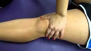
Palpation
- Medial/lateral epicondyle
- Olecranon & olecranon fossa
- Radial head
- Ulnar Collateral Ligament (UCL) of an elbow
Neurologic Assessment
- Reflexes: C5-C7
- Spinal Reflex/The Reflex Arc
- The reflex is the involuntary as well as nearly instantaneous movement in response to the stimulus. A reflex is an automatic response to the stimulus which does not receive or even need conscious thought as this happens through the reflex arc. Reflex arcs act on the impulse before that impulse reaches the brain.
- Reflex arcs can be
- Monosynaptic for example, contain only two neurons, the sensory, and the motor neurons. An instance of monosynaptic reflex arcs in humans includes a patellar reflex & an Achilles reflex.
- Polysynaptic for instance, multiple interneurons (also described as relay neurons) that interface between sensory along with motor neurons in a reflex pathway.
Technique for testing reflexes
- A muscle group to be tested can be in a normal (neutral) position (for example, neither stretched nor contracted).
- A tendon attached to a muscle(s) that is/are to be tested may be clearly identified. After that, place an extremity in the position which allows a tendon to be easily struck with a reflex hammer.
- Moreover, to easily locate a tendon, ask a patient to contract a muscle that is attached. When a muscle shortens, the person (the physical therapist) may be handled to both see as well as feel the cord namely the tendon, confirming it is precise location.
- Strike a tendon with a single, brisk, stroke. A physical therapist may not elicit pain.
The grading system is rather subjective
- 0: No evidence of contraction
- 1+: Reduced but still present (hypo-areflexia). Hyporeflexia is a generally associated with the lower motor neuron deficit (at alpha motor neurons from the spinal cord to muscle) for instance, Guillain–Barré syndrome
- 2+: Normal
- 3+: Super-normal (hyper-reflexes) Hyperreflexia is most often attributed to the upper motor neuron lesions, for example, Multiple sclerosis
- 4+ Clonus: Repetitive shortening of a muscle after a single stimulation to that muscle.
- Note any asymmetric increase or even depression. Jendrassik maneuver can be utilized to augment hypoactive reflexes for instance, the patient locks the hands together as well as pulls vigorously apart as the tendon in the lower extremity is tapped or can push the knees together against each other, whereas an upper limb tendon is tested.
Myotomes: C5-T1
Myotome
- The myotome (greek: myo=muscle, tome = a section, volume) is described as a group of muscles innervated by the single spinal nerve root. Myotome testing is an essential part of the neurological examination when suspecting radiculopathy. Myotomes are much more complex to test after dermatomes, each skeletal muscle is innervated by nerves derived from more than one spinal cord level.
- Myotomes are a part of the somatic nervous system as well as the somatic nervous system is a part of the peripheral nervous system.
Spinal Nerves
- There are thirty-one (31) spinal nerves, each vertebra has a spinal nerve. Nerves are categorized by the vertebra that houses them. There are eight(8) cervical nerves, twelve(12) thoracic nerves, five(5) lumbar nerves, five(5) sacral nerves, one(1) coccygeal nerve. Sixteen(16) of these thirty-one (31) nerves have a specific myotome that controls voluntary muscle movement.
Myotome Distribution
- Most muscles in the upper as well as limbs receive innervation from more than one spinal nerve root, as well as are hence comprised of multiple myotomes. For instance, the Biceps Brachii muscle flexes an elbow. This is innervated by a musculocutaneous nerve, that is innervated by C5, C6, and C7 nerve roots. All 3 of these spinal nerve roots may be said to be associated with elbow flexion.
The list below details which motion(s) has the strongest association with each myotome
- Upper Extremity:
C5- shoulder abduction.
C6– Elbow flexion Wrist extension
C7 – Elbow extension
C8 – Thumb Extension and wrist ulnar deviation
T1 – Finger abduction. - Chest wall and abdominal muscles:
T2 – L1[5] - Lower Extremity:
L2 – Hip flexion
L3 – Knee extension
L4 – Ankle dorsiflexion
L5 – Big toe extension
S1 – Ankle plantarflexion
S2 – Knee flexion
- Dermatomes: C5-T1
- Dermatomes
- The dermatome is the area of the skin that is mainly supplied by the single spinal nerve. There are eight(8) cervical nerves (note C1 has no dermatome), twelve(12) thoracic nerves, five(5) lumbar nerves as well as five(5) sacral nerves. Each spinal nerve relays sensation from a particular region of the skin to the brain.
- Dysfunction or even damage to the spinal nerve may trigger symptoms in the corresponding dermatome. In addition, nerve damage or dysfunction can result from infection, compression, or even traumatic injury.
- The nerves from the
- C2 to C4 supply the skin of a neck.
- C5 to T1 nerves supply the arms.
- T2 to L2 nerves supply the chest as well as the abdomen.
- L3 to S1 nerves supply the skin of the lower extremities(legs).
- S1 to S4 nerves go to the groin.
Movement Testing
- Active Range Of movement (AROM)/Passive Range Of Motion(PROM) with or even without overpressure
- Elbow
- Flexion
- Extension
- A positive Elbow Extension Test may indicate fracture along with the referral.
- Forearm pronation/supination
- Wrist
- Flexion
- Extension
- Cervical, Shoulder & Elbow Range of movement with or without overpressure
- Posterior-anterior glide assessment on the cervical/thoracic spine for distal symptoms reproduction as well.
- Resistive Testing
- Elbow flexion/extension
- Forearm pronation/supination
- Wrist flexion/extension
- Pain-free grip strength
- Thumb
- Fingers
- Accessory motion testing
- Humeroulnar traction
- Humeroradial traction
- Proximal/distal radioulnar Anterior/Posterior and Posterior/Anterior glides
Special Tests
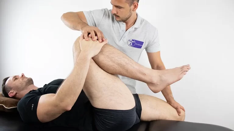
Elbow Flexion Test
- Purpose
- Elbow Flexion Test is a type of neurological dysfunction test that is utilized to determine a cubital tunnel syndrome (ulnar nerve ).
- Technique
- First of all, the patient should be in the position of standing or even sitting.
- After that, ask a patient to actively fully elbow flexion with the wrist extension as well as 90-degree shoulder girdle abduction & depression.
- Then, hold the position for up to two to three minutes.
- Next, a positive sign indicates feeling numbness or even tingling in the distribution of the ulnar nerve root.
Tinel’s Sign at Elbow
- Purpose
- Tinel’s Sign is the general term for a test in which the therapist identifies the irritated nerve through the percussive or even tapping technique. At an elbow, Tinel’s sign indicates the irritated Ulnar nerve.
- Technique
- The physiotherapist can be located the Ulnar nerve which is seated in the groove between the olecranon process as well as the medial epicondyle, the Ulnar nerve is then tapped repeatedly by the index finger of a physiotherapist. The positive sign is indicated by the tingling sensation in an ulnar distribution of the forearm and hand distal to a tipping point.
Ulnar Nerve Compression test
- For determining if a patient has ulnar nerve compression, the physician asks about the symptoms, takes the medical history, as well as does a complete examination of the arm, elbow, as well as hand. The physician can also test the arm for strength, sensation, as well as signs of nerve irritation or even damage.
- Grade (one)I: Mild symptoms include:
Intermittent paresthesia
Minor hypoesthesia of a dorsal as well as palmar surfaces of a fifth as well as medial aspect of the fourth digits
No motor changes - Grade (second)II: Moderate as well as persistent symptoms including:
- Paresthesia
Hypoesthesia of the dorsal as well as palmar surfaces of a fifth as well as medial aspect of the fourth digits
Mild weakness of the ulnar innervated muscles
Early signs of the muscular atrophy - Grade (three)III: Severe symptoms including:
- Paresthesia
Noticeable loss of the sensation of a dorsal as well as palmar surfaces of a fifth as well as medial aspect of the fourth digits.
Significant functional as well as motor impairment
Muscle atrophy of a hand intrinsic
Possible digital clawing of the fourth as well as fifth digits (Sign of Benediction) - Purpose
These tension tests are mainly done to check peripheral nerve compression or as part of the neurodynamic assessment. The main reason for using a ULTT is mainly for checking cervical radiculopathy. These tests are both diagnostic as well as therapeutic. Once a diagnosis of cervical radiculopathy is made the tests are completely done to mobilize an entrapped nerve. - Method
Each test is mainly performed on a normal/asymptomatic side first. Traditionally for an upper limb, the order of the elbow joint positioning is shoulder followed by the forearm, wrist, fingers, as well as lastly elbow. Each common joint positioning component is added till the pain is provoked or symptoms are reproduced. To further sensitivity upper limb tests, the side flexion of the cervical spine may be added. If the pain is provoked in a very starting position, after that there is no need to add further sensitizers.
Mill’s Test
- Purpose
The described test helps to diagnose Lateral Epicondylitis in an elbow, also known as “Tennis Elbow”. - Clinical presentation
The Patients report pain at a lateral elbow that radiates down a forearm. Moreover, the patients often complain of a weakened grip as well as difficulties lifting objects. On physical examination, the patients typically have point tenderness medial as well as distal to a lateral epicondyle. - Technique
- A Patient is in the sitting position as well as a clinician palpates a patient’s lateral epicondyle with one hand during pronating a patient’s forearm, fully flexing a wrist, and an elbow extended. The production of pain in an area of insertion at a lateral epicondyle indicates a positive test.
Cozen’s test
- Purpose
The purpose of Cozen’s test (also famous as a “resisted wrist extension test” or even “resistive tennis elbow test”) is to check for lateral epicondylalgia or even “tennis elbow”. - The patient position
A patient may be in the sitting position with an elbow extended forearm maximal pronation, the wrist radially abducted, as well as the hand in the fist. - An Examiner Position
A physiotherapist may stabilize an elbow during palpating the lateral epicondyle, the other hand placed on the dorsum of a hand.
Technique
A patient is asked to move a wrist to dorsal flexion as well as a physical therapist provides resistance to the motion, in a position described above. A test is positive if pain on a lateral epicondyle is elicited.
Maudsley’s test
- Purpose
Maudsley’s test is mainly utilized by clinicians to confirm a diagnosis of Lateral Epicondylitis ”Tennis Elbow”. - Epicondylitis represents the degenerative process involving an origin of extensor tendons at a lateral elbow as well as a flexor-pronator muscle group at a medial elbow. This is thought that repetitive stress, as well as overuse, lead to tendinosis with microtrauma as well as partial tearing that can progress to the full-thickness tendon tear. Lateral, as well as medial epicondylitis, are common disorders affecting the upper extremity. Epicondylitis elbow pain caused functional along with functional impairment moreover, typically results from specific occupational as well as sports-related motions. The Lateral epicondylitis, in the starting described by Morris as “lawn tennis elbow” in 1882 & now most commonly termed tennis elbow, can happen in patients while performing any activity that involves repeated supination as well as pronation of a forearm with an elbow in extension.
- Technique
The examiner resists extension of a 3rd digit of a hand, stressing an extensor digitorum muscle as well as tendon, during palpating a patient’s lateral epicondyle. Pain indicates a positive test over the lateral epicondyle of the humerus.
Chair Push-up Test
- Purpose
- A chair push-up test, also famous as the stand-up test or even chair sign, is utilized to test for a posterolateral rotatory instability (PLRI) of an elbow joint as well as an evaluation of its lateral collateral ligament.
- Technique
- A patient is in the sitting position in the chair with the two hands resting on a seat by the sides or a chair’s arms. A patient actively pushes up to transition into the standing position. Pain, apprehension, clicking, or even locking produced while a transition is indicative of posterolateral rotatory instability (PLRI) as well as the failure of the lateral collateral ligament.
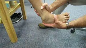
Elbow Varus Stress
- Purpose
- The main purpose of the varus stress test of the elbow is mainly done integrity of a lateral collateral ligament.
- Technique
- A patient is in the standing position, a physiotherapist places the patient’s elbow in slight flexion during palpating a humeroulnar joint line. A physiotherapist after that applies the varus force to an elbow. This described test is considered positive if a patient experiences pain or excessive laxity noted as well as compared to a contralateral side.
Elbow Valgus Stress
- Purpose
- An elbow valgus stress test is mainly utilized to assess the integrity of a medial collateral ligament, also famous as an ulnar collateral ligament.
- Technique
- The described test may be performed with a patient supine, sitting, or in a standing position. A physiotherapist places the patient’s elbow in approximately 20 degrees of the flexion during palpating a medial joint line as well as stabilizing a distal humerus with one hand as well as applying the valgus stress to an elbow with another hand. A described test is considered positive if a patient experiences pain or excessive laxity noted compared to a contralateral side. As with a varus stress test, the test may be repeated in varying degrees of elbow extension to test different portions of the medial collateral ligament(MCL).
Moving Valgus Stress Test
- Purpose
- A purpose of a moving valgus stress test is for assessing the integrity of a medial collateral ligament, or an ulnar collateral ligament of an elbow.
- Technique
- The described test may be done with a patient sitting or even standing. A physiotherapist abducts a patient’s shoulder to 90 degrees. A physiotherapist grasps a distal forearm with one hand as well as stabilizes an elbow with another. An examiner after that maximally flexes the elbow as well as places the valgus stress on an elbow during externally rotating a shoulder. When the shoulder reaches an end range of external rotation, an examiner quickly as well as smoothly extends an elbow to approximately 30 degrees.
- For this moving valgus stress test to be considered positive, 1) if a patient must experience pain at the medial elbow, and, 2) a maximal amount of pain may be experienced between 120 as well as 70 degrees of elbow flexion.
Neurodynamic Tests
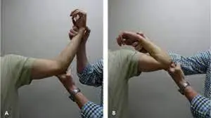
Upper Limb Tension Tests (ULTTs)
- Introduction
A Brachial Plexus Tension or Elvey Test, also famous as the Upper Limb Tension Test, are designed to put stress on the neurological structures of the upper limb by elongating them. These tests were first written by Elvey and are hence also famous as the Elvey test but most commonly called ULTT. A ULTT is equivalent to a straight leg raise designed for a lumbar spine. - Purpose
ULTT are done for assessment of the peripheral nerve mobility as well as compression or as part of the neurodynamic assessment. These tests are done as a cluster to make the confirmatory diagnosis for the nerve involvement. Once a diagnosis is made tests are done to mobilize an entrapped nerve. - Technique
ULTTs goal at evoking the patient’s symptoms. It is performed by keeping a shoulder, an elbow, a forearm, a wrist as well as fingers in specific positions to put stress on the particular nerve (nerve bias), that can be performed with modifications in the position of each joint as the “sensitizer” to the specific nerves. - General points which need to be kept in mind during doing a test.
- The physiotherapist may briefly explain to a patient a procedure they are going to do in layman’s terms. A reduces the feeling of anticipation among the patient, moreover, they are calmer as well as relaxed. In addition, inform the patient that it may or may not promote the symptoms as well as communicate verbally when a patient feels any kind of discomfort.
- Care may be taken to just evoke the symptoms as well as not further aggravate the symptoms.
- Each test is performed on a normal/asymptomatic side first.
- The precision of this test is attributed to doing it with the appropriate stabilizing hand as well as the slow, steady mobilizing hand.
The final action may not be held for more than seven to nine seconds. - Watch for unwanted motions, that might alter the results. Traditionally for an upper limb, the order of joint positioning is the shoulder followed by the forearm, the wrist, fingers, as well as lastly elbow. Each common positioning component is added till the pain is provoked or symptoms are reproduced. To further sensitize the upper limb test, side flexion of the cervical spine may be added. If the pain is provoked in a very starting position, there is no requirement to add further sensitizers.
- Positive Test
A described test is positive if one or more of the following occurs: - Symptoms reproduced
The side-to-side difference in the elbow extension is more significant than ten to fifteen degrees.
Contralateral cervical side bending increases symptoms, or even ipsilateral side bending reduces symptoms.
Upper Limb Tension Test (one)1 (ULTT1, for checking Median nerve compression)
- Indications:
Radiating pain in an upper limb,
Tingling sensations in first 3 fingers(thumb, index finger as well as middle finger). - Motions performed:
Shoulder depression,
Shoulder abduction 110 degrees,
Shoulder external rotation with an elbow is 90 degrees of flexion,
Forearm supination,
Wrist as well as finger extension,
Elbow extension. - Structural differentiation:
Proximal symptoms- Relieve wrist as well as a finger extension
Distal symptoms (provocation)- Contralateral flexion of the neck.
Upper Limb Tension Test 2A (ULTT2A, For checking Median nerve compression)
- Indications of this test:
Radiating pain in an upper limb,
Recent shoulder arthroplasty surgery,
Recent Dislocation of the shoulder as well as Instability. - Movements done:
Shoulder girdle depression,
Shoulder abduction 100 degrees,
External rotation with the elbow at 90 degrees,
Forearm supination,
Wrist as well as finger extension,
Elbow extension. - Structural differentiation:
Proximal symptoms: Relieve wrist as well as a finger extension
Distal symptoms (provocation): Contralateral flexion of the neck.
Upper Limb Tension Test 2B (ULTT2B, Radial nerve entrapment examination)
- Indications:
Radiating pain in an upper limb,
Supinator tunnel syndrome,
De Quervain`s disease,
Cervical Radiculopathy - Motions performed:
Shoulder girdle depression,
Shoulder abduction 20-30 degrees,
Shoulder internal rotation,
Forearm pronation,
Wrist, finger as well as thumb flexion,
Elbow extension. - Structural differentiation:
Proximal symptoms: Relieve wrist as well as finger flexion
Distal symptoms (provocation): Contralateral flexion of the neck
Upper Limb Tension Test 3 (ULTT3, checking Ulnar nerve compression)
- Indications:
Pain radiating to the 4th as well as 5th digits,
Thoracic outlet syndrome,
Carpal tunnel syndrome. - Movements done:
Shoulder girdle depression,
Shoulder abduction 110 degrees,
Shoulder external rotation,
Forearm pronation,
Wrist as well as Finger extension,
Elbow flexion. - Structural Differentiation:
Proximal symptoms: Relieve wrist as well as finger extension.
Distal symptoms (provocation): Contralateral flexion of the neck.
FAQ
Hold a forearm with one hand and, with an elbow flexed to 90°, palpate an elbow, feeling a head of a radius as well as a joint line with the thumb. If there is swelling present, is it fluctuant? Synovitis is usually felt as the fullness between an olecranon as well as a lateral epicondyle.
A normal arc is from zero degrees (full extension) to 135 degrees of flexion, as well as zero degrees to 180 degrees of rotation. When there is swelling or even pain localized to an elbow region, normal range of movement testing effectively rules out an elbow joint itself as a source of the issue.
This is essential to feel for crepitus as he moves an elbow joint (which may be associated with osteoarthritis) as well as observe any discomfort or even restriction in an elbow joint’s range of motion. If abnormalities are noted on the active motions(for instance, restricted range of movement), assess joint motions passively.
An elbow joint is the hinge joint that provides great stability as well as a motion for performing daily activities. Strong muscles that extend across an elbow joint bring about motions such as flexion, extension, supination as well as pronation enabling us to do activities of daily living.
Elbow joint
Bynovial folds of the elbow.
Bicipitoradial bursa.
Olecranon bursa as well as
Arcade of Struthers.
Ligament sprains, muscle strains, dislocation of the elbow, as well as osteoarthritis of an elbow joint. Bursitis (inflammation of a fluid-filled sac of an elbow joint).