Baxter’s Nerve Entrapment
Definition
Baxter’s neuropathy is a nerve entrapment syndrome that results from the compression of the inferior calcaneal nerve. Selective fatty atrophy of the abductor digiti minimi muscle is the unique sign of this neuropathy.
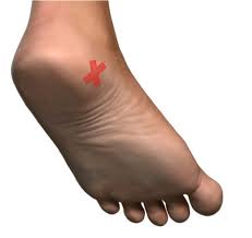
Baxter’s Entrapment is an entrapment (or compression) of the Inferior Calcaneal Nerve just under the base of the arch of the foot. The Inferior Calcaneal Nerve is the first branch of the Lateral Plantar Nerve on the bottom surface of the foot. The nerve is also sometimes called Baxter’s nerve, named after the first physician to describe this nerve entrapment as a specific cause of foot pain.
Table of Contents
Where is Baxter’s nerve?
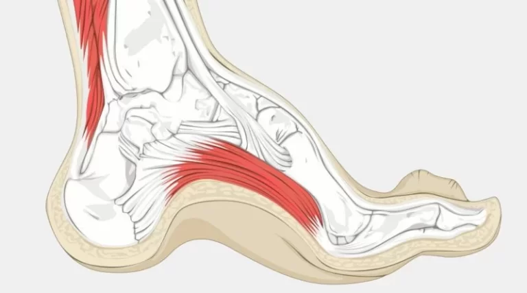
Baxter’s neuritis typically occurs where the nerve takes a right angle turn from the medial aspect of the calcaneus. It travels laterally across the plantar aspect of the foot to the abductor digiti minimi muscle anterior to the plantar tubercle of the calcaneus.
Causes-
One of the most common causes of chronic pain is entrapment of the first branch of the lateral plantar nerve, a condition that is known as Baxter’s neuropathy(17,18). It is believed that approximately 20% of cases of pain in the medial region of the heel are associated with neuropathy of that nerve
When one sees entrapment neuropathy of the Baxter’s nerve after a plantar fasciotomy, it is typically caused by distal migration of the fascia which can entrap the nerve or is the result of scar tissue which has bound the nerve down. Be aware that overzealous spur resection can also cause iatrogenic nerve damage
Symptoms

Patients will present with burning pain and paresthesia on the skin over the calcaneus. Pain does not radiate distally as in cases of the entrapment of the tibial nerve or Baxter’s nerve. Tinel’s sign might be positive over the medial surface of the calcaneal bone. When one sees entrapment neuropathy of the Baxter’s nerve after a plantar fasciotomy, it is typically caused by distal migration of the fascia which can entrap the nerve or is the result of scar tissue that has bound the nerve down.
The main reason why plantar fasciitis and Baxter’s nerve entrapment get confused is due to the similarity in the location of pain. In Baxter’s nerve entrapment, distinct tenderness is felt at the origin of the abductor hallucis muscle (a small muscle along the inside of the foot), whereas the most intense site of pain in plantar fasciitis is commonly more towards the bottom of the heel.
The main reason why plantar fasciitis and Baxter’s nerve entrapment get confused is due to the similarity in the location of pain. In Baxter’s nerve entrapment, distinct tenderness is felt at the origin of the abductor hallucis muscle (a small muscle along the inside of the foot), whereas the most intense site of pain in plantar fasciitis is commonly more towards the bottom of the heel.
Additionally, unlike plantar fasciitis, pain associated with entrapment tends to get worse with physical activity, rather than better. This is clinically known as post-kinetic dyskinesia. With entrapment, the pain is also more localised and is pressure sensitive. Consequently, in some cases, orthotic therapy may aggravate the pain if they have been prescribed to address a different diagnosis. This will be due to the orthotic further compressing the nerve. Other symptoms include burning and sharp shooting pain. Patients occasionally also locate the pain at the edges of the heel, either the outer or inner edge.
Risk Factors:
–Plantar Fasciitis
–Obesity
–Flat Foot
–Foot Hyperpronation
–Muscular Enlargement (such as in athletes)
–Bone Spur (Plantar Calcaneal Spur)
Diagnosing Baxter’s nerve entrapment
Due to the complexity of the foot and symptoms that are often indistinguishable to plantar fasciitis, diagnosing Baxter’s nerve entrapment requires a high degree of clinical suspicion and knowledge. Comprehensive history taking and clinical examination should be performed to assess for possible nerve related symptoms. This is extremely important as there is no definitive test to diagnose Baxter’s nerve entrapment. X-ray, ultrasound and MRI can be used, but only to rule out other possible conditions and risk factors. An effective diagnostic technique is to inject local anaesthetic to numb the possible offending nerve. If symptom relief is gained with the injection, it provides good evidence that Baxter’s nerve entrapment is present. Our podiatrists at OnePointHealth are able to perform this injection technique.
Conservative Treating Baxter’s nerve entrapment
Conservative treatment aims to reduce the causative factors of entrapment. Treatment may include:
Activity modification. An initial change to your exercise regime or daily activities may be needed to avoid the condition getting worse. The introduction of a low impact alternate activity may also be of benefit such as swimming.Icing: to reduce possible inflammation.Strengthening/Isometric loading. It is important that strength is addressed for this condition so that the affected area can tolerate more load. This is done initially with isometric loading to load up the affected area in a pain-free way.
Massage: to help release excessive tight soft tissue structures that surround the nerve.
Taping/splinting: to control poor foot motion and function.Orthotictherapy: can be used to address causative biomechanical factors such as flat feet, over-pronation.Injection therapy: local anaesthetic and corticosteroid can help bathe the nerve and reduce symptoms.
Some of the conservative treatment options are similar to those used for plantar fasciitis. However, with plantar fasciopathy pain levels tend to improve within weeks of a good treatment plan. In those who are non-responsive to conservative treatment, a diagnosis of Baxter’s nerve entrapment is further supported.
Surgery for Baxter’s nerve entrapment
Surgery is often indicated when pain levels fail to improve with conservative treatment. Additionally, if pain levels cease after an injection only for a short time before returning, surgery is also warranted. Surgery for Baxter’s nerve entrapment is known as neurolysis and may also be combined with a plantar fascia release.


One Comment