Test For Cervical Instability (Instability “Clearing” Tests)
Table of Contents
What is Test for Cervical Instability?
Testing for cervical instability involves a range of diagnostic exams and physical assessments to determine if the neck’s vertebrae and supporting structures are abnormally mobile or misaligned. These tests may include X-rays, MRI or CT scans, flexion-extension X-rays, and physical examinations. A healthcare professional may recommend testing for cervical instability if a patient is experiencing neck pain or other symptoms. Early detection and treatment of cervical instability can help prevent further damage and improve overall quality of life.
Description
Cervical spine instability is the most common condition that results from ligament damage. It is mainly transverse ligaments, alar ligaments, bone or joint damage, it may be dislocation or fracture, or weak muscles like deep flexors or extensors of cervical muscles.
The instability may result from chronic arthritic conditions like rheumatoid arthritis, trauma, long-term corticosteroid use, congenital malformation, down syndrome & osteoporosis. If in the history the patient complains of instability, a lump in the throat, lip paresthesia, severe headache (mostly with movement )muscle spasms, nausea, or vomiting, that leads high level of suspicion of instability
If the physiotherapist anticipates mobilizing mostly end-range techniques or manipulation techniques to the cervical spine, especially the upper cervical spine, a selection of appropriate clearing tests should be performed to rule out instability.
If any instability is present, mobilization and manipulation should not be performed.
Sign and Symptoms of Cervical instability
- Severe muscle spasm
- Lump in throat
- The patient does not want to move head (especially into the flexion position)
- Lip or facial paraesthesia
- Severe headache
- Dizziness
- Nausea
- Vomiting
- Nystagmus
- Soft end-feel
- Pupil changes
Cervical instability refers to the abnormal movement or excessive mobility of the cervical spine (neck). This condition may be caused by various factors such as trauma, congenital conditions, or degenerative changes in the spine.
Some of the common tests that may be used to diagnose cervical instability include:
- X-rays: These can provide images of the cervical spine and may reveal any abnormalities or misalignments.
- MRI, or magnetic resonance imaging, is a non-invasive diagnostic imaging test that uses powerful magnets and radio waves to create detailed images of the body’s internal structures.
- CT (computed tomography) scan: uses X-rays and computer technology to create detailed images of the cervical spine. A CT scan can show fractures or other abnormalities that may not be seen on X-rays.
- Flexion-extension X-rays: These are special X-rays that are taken while the patient moves their neck in different directions. This can help identify any abnormal movement or instability in the cervical spine.
- Physical examination: Your doctor may perform a physical examination to assess your neck’s range of motion, stability, and strength. They may also check for any neurological symptoms such as numbness, tingling, or weakness.
It’s important to consult with a healthcare professional if you have any concerns about cervical instability or neck pain. They can perform a thorough examination and recommend appropriate diagnostic tests or treatments.
Name of the test of Cervical instability
- Anterior shear stress test
- Lateral flexion alar ligament stress test
- Rotational alar ligament stress test
- Transverse ligament stress test
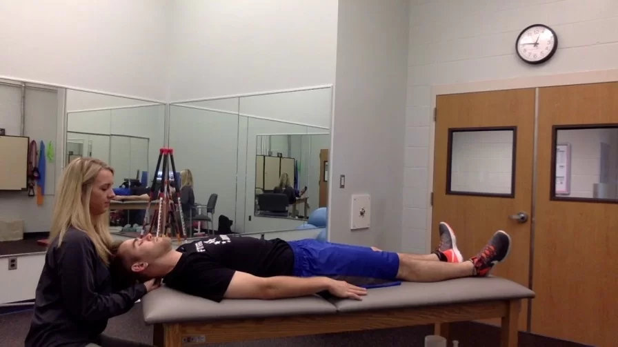
Anterior shear test.
This test is also called a sagittal stress test. Anterior shear stress is similar to the posteroanterior central vertebral pressure (PACVP) testing in the joint play section.
Importance: This test is planned to test the probability of the supporting ligaments and capsular tissues of the cervical spine.
Technique=
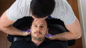
- The patient lies supine with their head in neutral resting on the bed.
- The physiotherapist applies an anterior direct pressure via the posterior arch of C1 or spinous processes of C2 to T1 or bilaterally through the lamina of all vertebral bodies.
- In this case, the normal end feel is tissue stretch
Result = When the upper cervical spine is performed, symptoms such as pupil changes, nystagmus, dizziness, soft end feel, nausea, facial or lip paresthesia, & a lump sensation in the throat are present the This test is Positive.
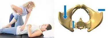
Lateral flexion Alar ligament stress test.

Purpose = This test is used to check the end range of motion (end feel) and capsular instability of the cervical spine.
Technique =
- The patient lies in a supine position with the head in the physiologically neutral position.
- while the physiotherapist stabilizes the axis with a wide pinch grip around the lamina and spinous process.
- The physiotherapist then attempts to side-flex the head and axis.
Result = Basically, if the ligament is intact, that leads to minimal side flexion occurs with a strong capsular end feel & a solid stop.
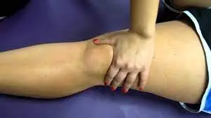
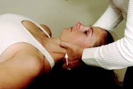
Lateral (transverse ) shear test.
Importance = Transverse shear test is used to identify the instability of the atlantoaxial articulation caused by odontoid dysplasia.
Technique =
- The patient lies supine with the head sustained.
- The examiner places the radial side of the second MCP joint of one hand against the transverse process of the atlas and the metacarpophalangeal (MCP) joint of the other hand against the contrast transverse process of the axis.
- The physiotherapist’s hands are then carefully pushed together, which leads to a shear force of one bone on the other bone.
- Normally, minimal motion & no symptoms appeared.
Result = This test is generally painful due to the compression of soft tissues as opposed to the bone, the patient should be warned the pain is a normal sensation to be expected.
This test can also be used to test different levels of the cervical spine ( C2 to C7 ).
Rotational Alar ligaments stress test:-
Importance = The transverse alar ligament stress test is used to identify the instability of the cervical spine.
Rotational Alar ligaments stress test
Technique =
Position of the patient is sitting position.
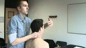
The examiner (therapist) grasps the lamina and spinous process of C2 in between the finger & thumb.
While stabilizing C2, the examiner ( therapist ) passively rotates the patient’s head to the left or right side and moves to the “no-symptom” side first.
If more than 20 degrees to 30 degrees of rotation is possible without C2 moving, it indicates injury to the contralateral Alar ligament particularly if the lateral bending alar stress test is positive in the same direction.
If they make the exaggerated motion in different directions for both tests, the cervical instability is because of an increase in the neutral zone in the joint of the cervical spine.
Kaale et al. advocated doing that test in a different fashion.
They supported placing the patient in a sitting position.
The examiner ( therapist ) stabilized the patient’s head instead of his or her body and placed both hands on the same side of the patient’s occipital-cervical junction.
The lower hand stabilized C2 by shoving the second and third fingers against the lateral aspect of C2 pulling its reward.
Then another hand is placed over the third finger under the lateral mass of the atlas and the second finger under the mastoid process pulls upward into rotation.
This test is taken out at the various angles of rotation that locate the position of greatest movements between C1 & C2.
FAQ
it may be difficult to hold up your head for a long period of time. it feels like heaviness in the head. Pain is present in the upper neck near the base of the skull. Pain referred to the shoulder.
Interpretation: Magnetic resonance imaging(MRI) is sensitive to soft-tissue injuries of the cervical spine. When a CT scan and radiography find no fractures or any types of instability, MR imaging does not help in deciding cervical stability and it may lead to unnecessary testing when not otherwise indicated.
Physiotherapy is useful for cervical instability.
Chiropractic.
Surgery.
Strengthening Exercises are helpful for cervical instability.
It is a medical condition in which loose ligaments in the upper cervical spine, may lead to damage of neurological structures and seen unacceptable symptoms. If they have cervical instability present, they may be experiencing migraines, vertigo, or nausea. providentially, cervical mobility is treatable.
Genetic dysfunctions or conditions: There are a handful of syndromes that cervical instability a related condition, including conditions like Down Syndrome, and rheumatoid arthritis (RA): In rheumatoid arthritis (RA) and similar to any arthritic conditions, they lead to increasing worsening of the joints and vertebrae can lead to cervical instability.