TENSOR FASCIA LATA TIGHTNESS :
Table of Contents
Introduction :
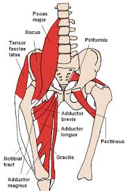
- When the Tensor fasciae latae tightness (TFL) is tight and locked up, it may cause pain in the hip joint, groin, buttock, lower back (over sacroiliac joint) and even the lateral aspect of the thigh.
- The Tensor fascia latae Tightness is most common seen in adult and stretching exercise is very important for keep your posture correct and also helps in reducing pain in Hip and Knee pain.
- Tensor fascia latae Tightness will create added tension through the iliotibial band (ITB).
- This increased tension in the ITB can create pain and irritation in the outer region of the knee, known as ITB syndrome.
- Tightness of TFL is also common mostly sportman involved in horse riding, running, and cycling.
- The Tensor fascia latae (TFL) is a fusiform muscle enclosed between two layers of fascia lata with a length of 15cm approximately and overlying the gluteus minimus and some part of the gluteus medius .
- It’s myotomes is fourth lumbar nerve root (L4)
ANATOMY OF TFL MUSCLE :
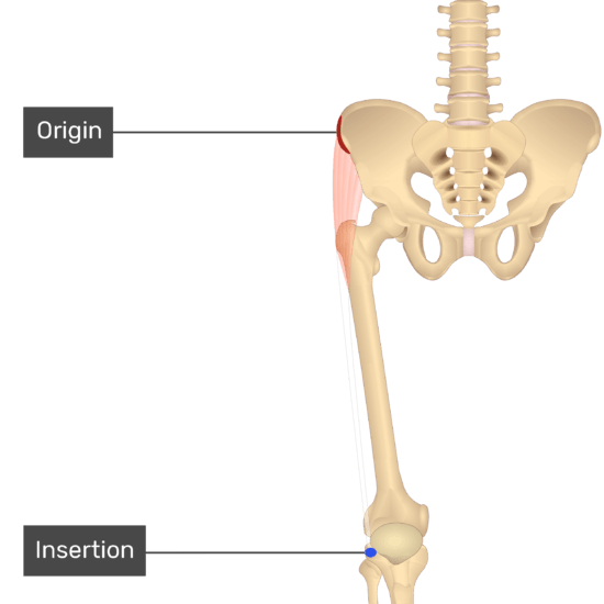
Origin:
- TFL originates from anterior superior iliac spine(ASIS) and the anterior part of iliac cres
Insertion :
- TFL together with the gluteus maximus joins to form the Iliotibial tract, which attaches to lateral condyle of tibia
- The iliotibial band is a 5 cm wide band called that divides into two layers superiorly and a single tract inferiorly. The superficial part is attached to the tubercle of the iliac crest, and the deep lamina to the capsule of the hip joint.
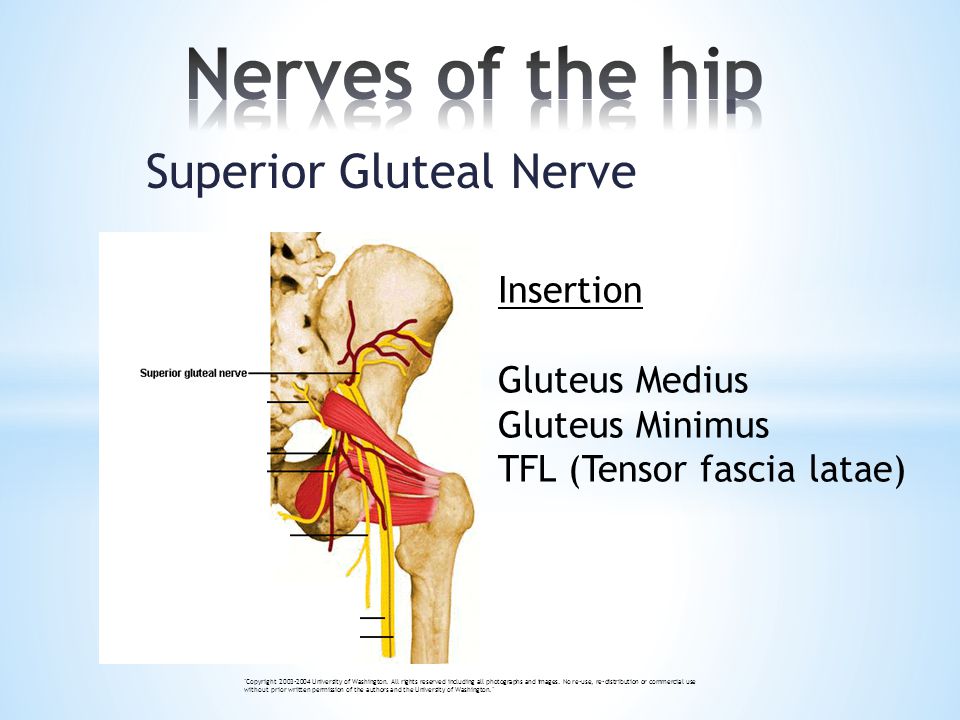
Nerve Supply :
- The Tensor fasciae latae is innervated by superior gluteal nerve, originating from lumbar nerve 4, 5, and first sacral nerve(L4-S1) roots. It also innervates gluteus minimus and medius muscles before terminating with innervation of tensor fasciae latae muscle.
Blood Supply :
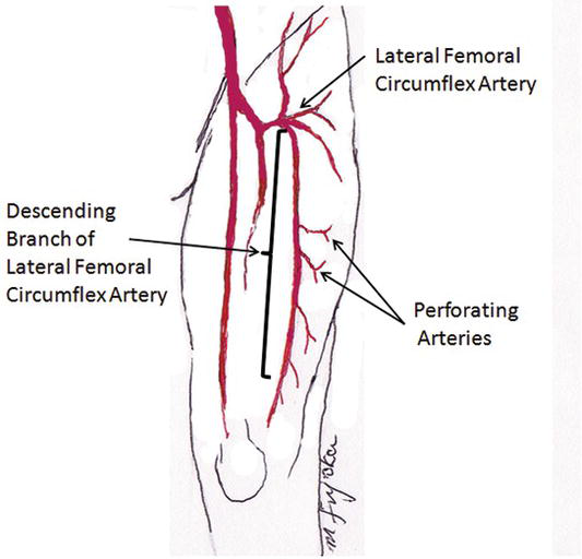
- Blood is supplied to TFL via deep branch of superior gluteal artery
- Ascending branch of the lateral circumflex femoral (LCF) artery
- The superior gluteal vien a branch of the internal iliac viens serves as the venous drainage route.
Function :
- TFL is prime mover in hip medial rotation and a weak hip abductor
- It serves as an accessory muscle/ hip synergist in abduction and flexion of the hip
- Together with gluteus maximus and the illiotibal band, it stabilizes the hip joint by holding the head of the femur in the acetabulum
- The tensor of fasciae latae, together with the gluteus maximus contributes in the stability of the knee during extension and also in partial flexion
- As part of the iliotibial tract it aids lateral rotation of the leg.
- TFL also assist in walking by inferiorly tilting the ilium on the weight-bearing side, with result in an upward tilt of the contralateral hip. Thus, allowing the leg of the non -weight bearing hip to swing through without hitting the ground during the swing phase of the gait.
Which are the Causes of Tensor fasciae latae tightness :
There are too many cause of TFL Tightness, Few most common reason are as per below :
- Sedentary Life Style
- Office Worker : If You are doing office Job then your TFL muscle may be Tight.
- Over-striding : It is running with a stride that is longer than optimal for your body length.
- Hip Pain due to Injury.
- Running on crowned roads
- Bow legs for any reasons.
- Shoe factor : worn out running shoes or shoes without beefed up heels.
- The main cause for TFL pain is overuse and compensation for weaker surrounding muscles. Pain occurring in muscles is often the result of the muscles compensating or working much harder than they were made to work. This compensation occurs due to surrounding muscles being dysfunctional due to inhibition or weakness.
Tensor fasciae latae tightness symptoms :
Tight TFL cause back pain
- And when we stand up, if it’s real tight, the muscle will pull us into kind of what we call an anterior pelvic tilt, which can compress the low back. There are many things that can be caused by tight TFL.
Cycle with TFL pain
- You may find that you have recurring knee or hip pain each year or develop it after cross-training or running. A tight TFL alone may not be problematic but when mixed with bike fit problems it is a season ending injury waiting to happen. Traditionally, the TFL is ignored when it comes to cycling, it is time that stops.
Tight TfL cause groin pain
- IT can include hip, groin or outer knee pain that feels sharp,
- stabbing or burning,
- as well as a click or pop. Usually,
- symptoms onset gradually then worsen with continued running or other repetitive activities.
Tight TFL causing Knee pain :
- When the TFL is tight, it can contribute to knee pain and injuries such as the iliotibial band syndrome (ITBS), patellofemoral pain syndrome (PFPS), and even meniscus injuries. Running shortens the TFL muscle because the hip is continually contracting with each stride.
Assessment :
Power :
- The power test for the TFL is done in side-lying with hip in 45° of flexion for grade 5, 4 and 3 while for grade 2, 1 and 0, it is done in long sitting position.
- The therapist is situated behind the patient with one hand is placed at the lateral surfece of the topmost thigh immediately above the knee, given downwards pressure and the other hand at the iliac crest for stability. the patient is asked to abduct against resistance.
- Grade 5 is given if the patient is able to abduct and maintain position at end range against maximal resistance.
- Grade 4 is given if the patient is able to abduct and maintain position at end range against moderate resistance.
- Grade 3 is given if the patient is able to abduct and maintain position at end range against no resistance except gravity.
- The therapist stands at the side of the limb being assessed one hand is place at the ankle serving to reduced friction between the limb and the plinth while the other hand is placed at the proximal anterolateral thigh. The patient is asked to move the limb towards the therapist.
- Grade 3 is given if the patient is able to abduct to 30° and maintain position at end range.
- For grade 1 and 0, the starting position is the same for grade 3 assessment but the hand placement of the therapist changes;one hand is placed at the the lateral side of the thigh immediately above the knee while the other hand is placed at the proximal anterolateral thigh for ease of TFL palpation.The patient is asked to move the limb towards the therapist.
- Grade 1 is given if therapist is able to feel contraction of the muscle and grade 0 is given if no contraction is palpable.
Ober Test :
- Frank Ober described the test for tightness of TFL and ITB in an article entitled “Back Strain and Sciatica”, wherein he discussed the relationship of a contracted TFL and ITB to low backache .
Test position :

- The patient should be in side-lying with the affected side up.
- Bottom knee and hip should be flexed to flatten the lumbar curve.
- Stand behind the patient and firmly stabilize the pelvis/greater trochanter to prevent movement in any direction.
- Grasp the distal end of the patient’s affected leg with your other hand and flex the leg to a right angle at the knee
Test:
- Extend and abduct the hip joint.
- Slowly lower the leg toward the table -adduct hip- until motion is restricted.
- Ensure that the hip does not internally rotate and flex during the test and the pelvis must be stabilized. As allowing the thigh to drop in flexion and internal rotation would ‘give in’ to the tight TFL and not accurately test the length.
Results:

- If the ITB is normal, the leg will adduct with the thigh dropping down slightly below the horizontal and the patient won’t experience any pain; in this case, the test is called negative.
- If the ITB is tight, the leg would remain in the abducted position and the patient would experience lateral knee pain, in this case, the test is called positive.
- Modified Ober Test[5]
- A modification of the Ober test was first recommended by Kendalls in Posture and Pain. In this test, there is less strain on the medial aspect of the knee joint, less tension on the patella, less interference by a tight Rectus Femoris and it provides a complete stretch on TFL.
Test position:
- The patient is in a side-lying position with underneath leg flexed at hip and knee to flatten the low back.
- The examiner stabilizes the pelvis and keeps the lateral trunk in contact with the table.
- Note: Downward lateral tilt is equivalent to the hip abduction and that would “give in ” to a tight TFL.
- Test:
- The examiner extends the leg. Ensure that leg is not internally rotated.
Results:
- With the knee straight and the pelvis in a neutral position, the thigh drops about 10 degrees below the horizontal. It is suggestive of normal length.
Palpation :
- Palpation of TFL can be done in any of the aforementioned starting position in muscle power testing. One hand is placed at the the lateral side of the thigh immediately above the knee while the other hand is placed at the proximal anterolateral thigh and the patient is instructed to abduct the limb being assessed.
- TFL can then be easily palpable at end range of the motion
CLINICAL CONDITION :
ITB syndrome :
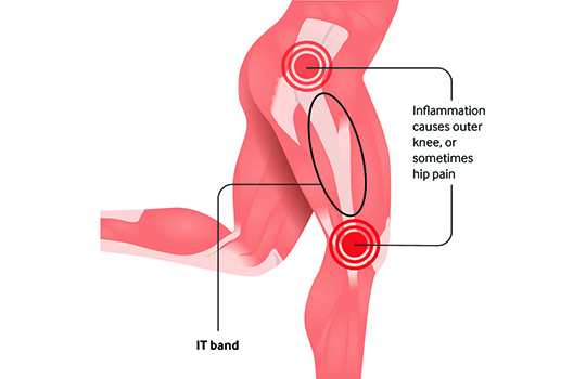
- Iliotibial band syndrome (ITBS or IT band syndrome) is an overuse injury of the connective tissues that are located on the lateral or outer part of thigh and knee.
- It causes pain and tenderness in those areas, especially just above the knee joint.
PHYSIOTHERAPY TREATMENT :
PAIN RELIVING :

- Rest, ice, compression, and elevation (RICE Principle).
- Anti-inflammatory medications, like ibuprofen (Advil, Motrin) and naproxen (Aleve)
- Physiotherapy may be needed to decrease the inflammation at the IT band.
- Ultrasound Mdalities may be used, including phonopheresis and iontophoresis to help decrease irritation in the soft tissues surrounding the knee.
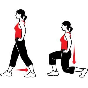
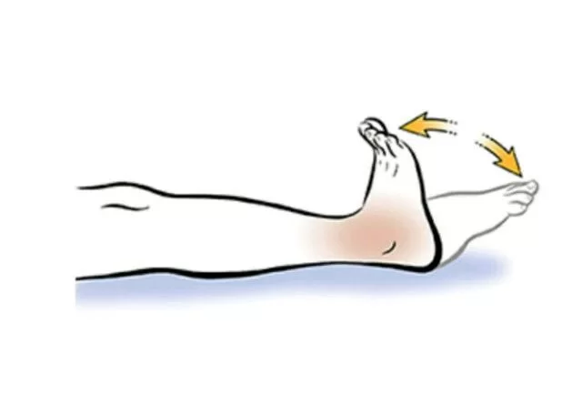
How to stretch the tensor fasciae latae ?
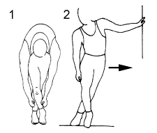
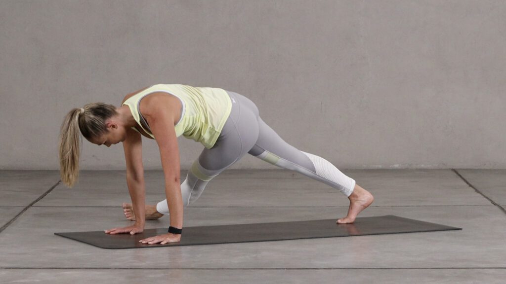
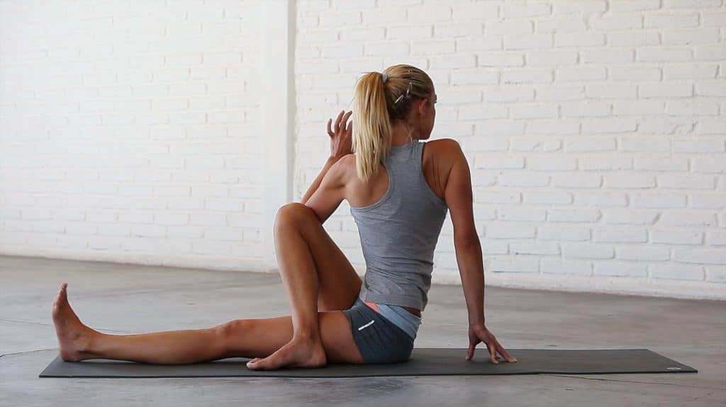
TFL STRETCHING EXERCISE :
STRETCH 1 OF TFL MUSCLES
STRETCH 2
STRETCH 3

STRETCH 4
STRETCH 5


2 Comments